File:Hertig1956 fig51.jpg

Original file (712 × 1,095 pixels, file size: 109 KB, MIME type: image/jpeg)
Plate 9
Two villous ova of 16 to 17 days of age. Streeter Horizon VII encompasses that developmental stage characterized by branching villi and an axis of the germ disc, whereas Horizon VIII includes that stage characterized by Hensen’s node and primitive groove. The younger of these two ova has not only branching villi and a well-defined axis of the germ disc, but also a definite primitive streak (although not a very distinct primitive groove) and the first beginnings of a head process. Heuser et. al. when reporting this specimen in detail (K15) observed the apparent overlapping of the criteria listed for Horizon VII. and VIII, and considered the younger specimen (7802) an advanced member of Horizon VII. The embryo of the older specimen as yet has not been studied in detail and ultimately may well be placed at least. in Horizon VIII. in this publication our primary interest is in the extra-embryonic structures of these ova.
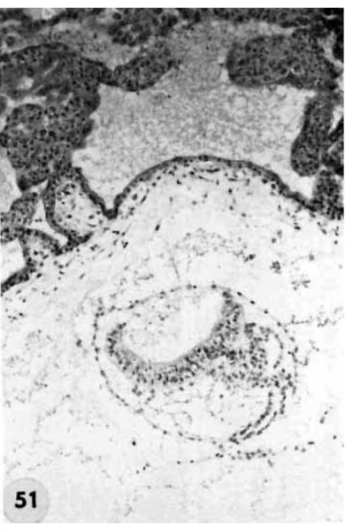
51 A medium-power view of a section of the ovum illustrated in figure 50 to show detail of the embryo. Note that the amnion has increased in size and is enfolding the germ disc and that. there is increased differentiation of the ectoderm and cndoderm at the right. The stems of two villi seen at the upper left have a mesoblastic core covered by inner Langhans epithelium (cytotrophoblast) and an outer thin layer of syncytium. Early angiogenesis is seen in the mesoblast of these villi t'oresha«lowing the development of the future fetal circulation which will be completed and functioning in 4 or 5 days (7-17 somites). During this period of time the amount of blood circulating through the spiral artcriolar sinusoids, the intervillous space and the venous sinusoids of the utero-placental circulation will also increase. Thus are establishcd the two contiguous circulations, separated only by the trophoblast, fibrous tissue and endothelium — the placental barrier. By this mechanism there results an interchange of nutritive, waste and other substances between the embryo and mother. Carnegie 8602, Section 25-3-2. X 100.
- Figure Links: 1 | 2 | 3 | 4 | 5 | 6 | 7 | 8 | 9-10 | 11-12 | 13-14 | 15-16 | 17 | 18-19 | 20 | 21-22 | 23 | 24-25 | 26-27 | 28-29 | 30-31 | 32-33 | 34 | 35 | 36 | 37 | 38 | 39 | 40 | 41 | 42 | 43 | 44 | 45 | 46 | 47 | 48 | 40 | 49 | 50 | 51 | 52 | 53 | 54 | 55 | 56 | 57 | 58 | 59 | 60 | 61 | 62 | 63 | 64 | 65 | 66 | 67 | 68 | 69 | 70 | 71 | 72 | 73 | 74 | 75 | 76 | 77 | 78 | 79 | 80 | 81 | 82 | 83 | 84 | 85 | 86 | 87 | 88 | 89 | 90 | plate 1 | plate 2 | plate 3 | plate 4 | plate 5 | plate 6 | plate 7 | plate 8 | plate 9 | plate 10 | plate 11 | plate 12 | plate 13 | plate 14 | plate 15 | plate 16 | plate 17 | table 1 | table 1 image | table 2 image | table 3 image | table 4 | table 4 image | table 5 | table 5 image | All figures | 1956 Hertig | Embryology History - Arthur Hertig | John Rock | Historic Papers
Reference
Hertig AT. Rock J. and Adams EC. A description of 34 human ova within the first 17 days of development. (1956) Amer. J Anat., 98:435-493.
Cite this page: Hill, M.A. (2024, April 27) Embryology Hertig1956 fig51.jpg. Retrieved from https://embryology.med.unsw.edu.au/embryology/index.php/File:Hertig1956_fig51.jpg
- © Dr Mark Hill 2024, UNSW Embryology ISBN: 978 0 7334 2609 4 - UNSW CRICOS Provider Code No. 00098G
File history
Click on a date/time to view the file as it appeared at that time.
| Date/Time | Thumbnail | Dimensions | User | Comment | |
|---|---|---|---|---|---|
| current | 22:16, 23 February 2017 | | 712 × 1,095 (109 KB) | Z8600021 (talk | contribs) |
You cannot overwrite this file.
File usage
The following 2 pages use this file: