File:Hertig1956 fig28-29.jpg: Difference between revisions
No edit summary |
m (→Fig. 28-29.) |
||
| (One intermediate revision by the same user not shown) | |||
| Line 1: | Line 1: | ||
==Fig. 28-29.== | |||
Three normal, late previllous ova 11 to 12 days old, of Horizon Vc. characterized by intereommunieating laeunar spaces filled with maternal plasma containing still increasing numbers of blood cells, a distended chorionic. cavity, an execoelomic cavity of maximum size, a bilaminar germ disc and well~defined amnion and amniotic cavity. Details of the germ discs of these ova are shown on plate 6. | |||
28 A surface view of an intact 12-day ovum after fixation, dehydration and clearing to show the appearance of maternal blood in the lacunar spaces. This is the most distinctive gross feature of, and first. appears at, this stage of develop ment. It is due to flooding of the trophoblastic. lacunae with maternal blood. [[:Category:Carnegie Embryo 7700|Carnegie 7700]], Sequence 9. X 22. | |||
29 A medilun-power view of a. mid-cross section of the ovum shown in figure 28. Early decidua is developing in the area iimnediately surrounding the ovum. Note three irregular masses of cytotrophohlast-—primordial villi—projccting into the syncytium. Note fully developed cxocoelomic cavity and membrane with a loose. mesh-work of mesohlasts between it and the cytotroplioblast lining the «ho:-ionic cavity. The trophoblast at the abembryonic pole is poorly differentiated, as contrasted to that at the embryonic or implantation pole. [[:Category:Carnegie Embryo 7700|Carnegie 7700]], Section 6-1-5. X100. | |||
{{Hertig1956 figures}} | |||
[[Category:Carnegie Embryo 7700]][[Category:Carnegie Stage 5]] | |||
Latest revision as of 10:43, 24 February 2017
Fig. 28-29.
Three normal, late previllous ova 11 to 12 days old, of Horizon Vc. characterized by intereommunieating laeunar spaces filled with maternal plasma containing still increasing numbers of blood cells, a distended chorionic. cavity, an execoelomic cavity of maximum size, a bilaminar germ disc and well~defined amnion and amniotic cavity. Details of the germ discs of these ova are shown on plate 6.
28 A surface view of an intact 12-day ovum after fixation, dehydration and clearing to show the appearance of maternal blood in the lacunar spaces. This is the most distinctive gross feature of, and first. appears at, this stage of develop ment. It is due to flooding of the trophoblastic. lacunae with maternal blood. Carnegie 7700, Sequence 9. X 22.
29 A medilun-power view of a. mid-cross section of the ovum shown in figure 28. Early decidua is developing in the area iimnediately surrounding the ovum. Note three irregular masses of cytotrophohlast-—primordial villi—projccting into the syncytium. Note fully developed cxocoelomic cavity and membrane with a loose. mesh-work of mesohlasts between it and the cytotroplioblast lining the «ho:-ionic cavity. The trophoblast at the abembryonic pole is poorly differentiated, as contrasted to that at the embryonic or implantation pole. Carnegie 7700, Section 6-1-5. X100.
- Figure Links: 1 | 2 | 3 | 4 | 5 | 6 | 7 | 8 | 9-10 | 11-12 | 13-14 | 15-16 | 17 | 18-19 | 20 | 21-22 | 23 | 24-25 | 26-27 | 28-29 | 30-31 | 32-33 | 34 | 35 | 36 | 37 | 38 | 39 | 40 | 41 | 42 | 43 | 44 | 45 | 46 | 47 | 48 | 40 | 49 | 50 | 51 | 52 | 53 | 54 | 55 | 56 | 57 | 58 | 59 | 60 | 61 | 62 | 63 | 64 | 65 | 66 | 67 | 68 | 69 | 70 | 71 | 72 | 73 | 74 | 75 | 76 | 77 | 78 | 79 | 80 | 81 | 82 | 83 | 84 | 85 | 86 | 87 | 88 | 89 | 90 | plate 1 | plate 2 | plate 3 | plate 4 | plate 5 | plate 6 | plate 7 | plate 8 | plate 9 | plate 10 | plate 11 | plate 12 | plate 13 | plate 14 | plate 15 | plate 16 | plate 17 | table 1 | table 1 image | table 2 image | table 3 image | table 4 | table 4 image | table 5 | table 5 image | All figures | 1956 Hertig | Embryology History - Arthur Hertig | John Rock | Historic Papers
Reference
Hertig AT. Rock J. and Adams EC. A description of 34 human ova within the first 17 days of development. (1956) Amer. J Anat., 98:435-493.
Cite this page: Hill, M.A. (2024, April 26) Embryology Hertig1956 fig28-29.jpg. Retrieved from https://embryology.med.unsw.edu.au/embryology/index.php/File:Hertig1956_fig28-29.jpg
- © Dr Mark Hill 2024, UNSW Embryology ISBN: 978 0 7334 2609 4 - UNSW CRICOS Provider Code No. 00098G
File history
Click on a date/time to view the file as it appeared at that time.
| Date/Time | Thumbnail | Dimensions | User | Comment | |
|---|---|---|---|---|---|
| current | 20:44, 23 February 2017 | 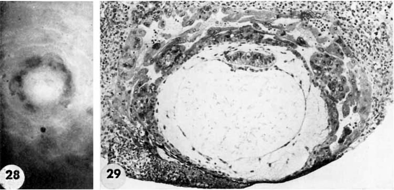 | 1,280 × 619 (139 KB) | Z8600021 (talk | contribs) |
You cannot overwrite this file.
File usage
The following 3 pages use this file: