File:Hertig1956 fig04.jpg
Hertig1956_fig04.jpg (745 × 562 pixels, file size: 41 KB, MIME type: image/jpeg)
Fig. 4.
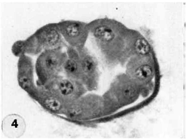
Normal preimplantation stages in human development; one segmenting egg found in the tube, Streeter Horizon II, and two free intrauterine blastocysts, Streeter Horizon III.
4 Mid-serial section of 58-cell intrauterine blastocyst shown as intact specimen in figure 3. Note absence of zona, about two-thirds of the periphery, probably of artifactitious origin. The two polar bodies are evident at opposite poles of the ovum. The inner cell mass of 5 blastomeres, only 4 of which are present in this section, is lying eccentrically within the segmentation cavity and surrounded by primitive troplioblastic blastomcres. The primitive segmentation cavity, composed of several coalescing spaces, is most clearly delineated as a single unit in this and the two contiguous sections. Note that the nuclei of the embryonic blastomeres, although of equal size, are less vesicular and contain denser clumps of chromatin than those of the trophoblastic blastomeres. Carnegie 8794, Section 7. X 500.
- Figure Links: 1 | 2 | 3 | 4 | 5 | 6 | 7 | 8 | 9-10 | 11-12 | 13-14 | 15-16 | 17 | 18-19 | 20 | 21-22 | 23 | 24-25 | 26-27 | 28-29 | 30-31 | 32-33 | 34 | 35 | 36 | 37 | 38 | 39 | 40 | 41 | 42 | 43 | 44 | 45 | 46 | 47 | 48 | 40 | 49 | 50 | 51 | 52 | 53 | 54 | 55 | 56 | 57 | 58 | 59 | 60 | 61 | 62 | 63 | 64 | 65 | 66 | 67 | 68 | 69 | 70 | 71 | 72 | 73 | 74 | 75 | 76 | 77 | 78 | 79 | 80 | 81 | 82 | 83 | 84 | 85 | 86 | 87 | 88 | 89 | 90 | plate 1 | plate 2 | plate 3 | plate 4 | plate 5 | plate 6 | plate 7 | plate 8 | plate 9 | plate 10 | plate 11 | plate 12 | plate 13 | plate 14 | plate 15 | plate 16 | plate 17 | table 1 | table 1 image | table 2 image | table 3 image | table 4 | table 4 image | table 5 | table 5 image | All figures | 1956 Hertig | Embryology History - Arthur Hertig | John Rock | Historic Papers
Reference
Hertig AT. Rock J. and Adams EC. A description of 34 human ova within the first 17 days of development. (1956) Amer. J Anat., 98:435-493.
Cite this page: Hill, M.A. (2024, April 27) Embryology Hertig1956 fig04.jpg. Retrieved from https://embryology.med.unsw.edu.au/embryology/index.php/File:Hertig1956_fig04.jpg
- © Dr Mark Hill 2024, UNSW Embryology ISBN: 978 0 7334 2609 4 - UNSW CRICOS Provider Code No. 00098G
File history
Click on a date/time to view the file as it appeared at that time.
| Date/Time | Thumbnail | Dimensions | User | Comment | |
|---|---|---|---|---|---|
| current | 19:27, 23 February 2017 |  | 745 × 562 (41 KB) | Z8600021 (talk | contribs) |
You cannot overwrite this file.
File usage
The following 2 pages use this file:
