File:Hertig1946b fig26.jpg

Original file (800 × 1,299 pixels, file size: 290 KB, MIME type: image/jpeg)
Fig. 26. A human placenta at term
To show the cystic degeneration occurring in the “fibrinoid" of a septum separating two adjacent cotyledons. B. L.-i. H., S-34-581.
A. A low power view of two cotyledons separated by a cytotrophoblastic septum whose "fibrinoid” has undergone cystic degeneration. The resultant hematoma at the base has disrupted the blood supply of the cotyledon at the left and caused infarction but has spared the one to the right. X5.
B. A higher powered view of the rectangular area seen in A. Note degenerated septum with recent hemorrhage below. The villi at the left are congested and undergoing early necrosis whereas the ones at the right are normal. X60.
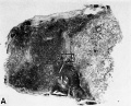
26A
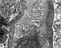
26B


References
Hertig AT. lnvolution of tissues in fetal life: a review. (1946) Anat. Rec. 94: 96-116.
Cite this page: Hill, M.A. (2024, April 27) Embryology Hertig1946b fig26.jpg. Retrieved from https://embryology.med.unsw.edu.au/embryology/index.php/File:Hertig1946b_fig26.jpg
- © Dr Mark Hill 2024, UNSW Embryology ISBN: 978 0 7334 2609 4 - UNSW CRICOS Provider Code No. 00098G
File history
Click on a date/time to view the file as it appeared at that time.
| Date/Time | Thumbnail | Dimensions | User | Comment | |
|---|---|---|---|---|---|
| current | 10:46, 8 August 2017 | | 800 × 1,299 (290 KB) | Z8600021 (talk | contribs) | ==Fig. 26. A human placenta at term== To show the cystic degeneration occurring in the “fibrinoid" of a septum separating two adjacent cotyledons. B. L.-i. H., S-34-581. A. A low power view of two cotyledons separated by a cytotrophoblastic septum... |
You cannot overwrite this file.
File usage
The following page uses this file: