File:Hertig1946b fig15.jpg
Original file (800 × 1,348 pixels, file size: 196 KB, MIME type: image/jpeg)
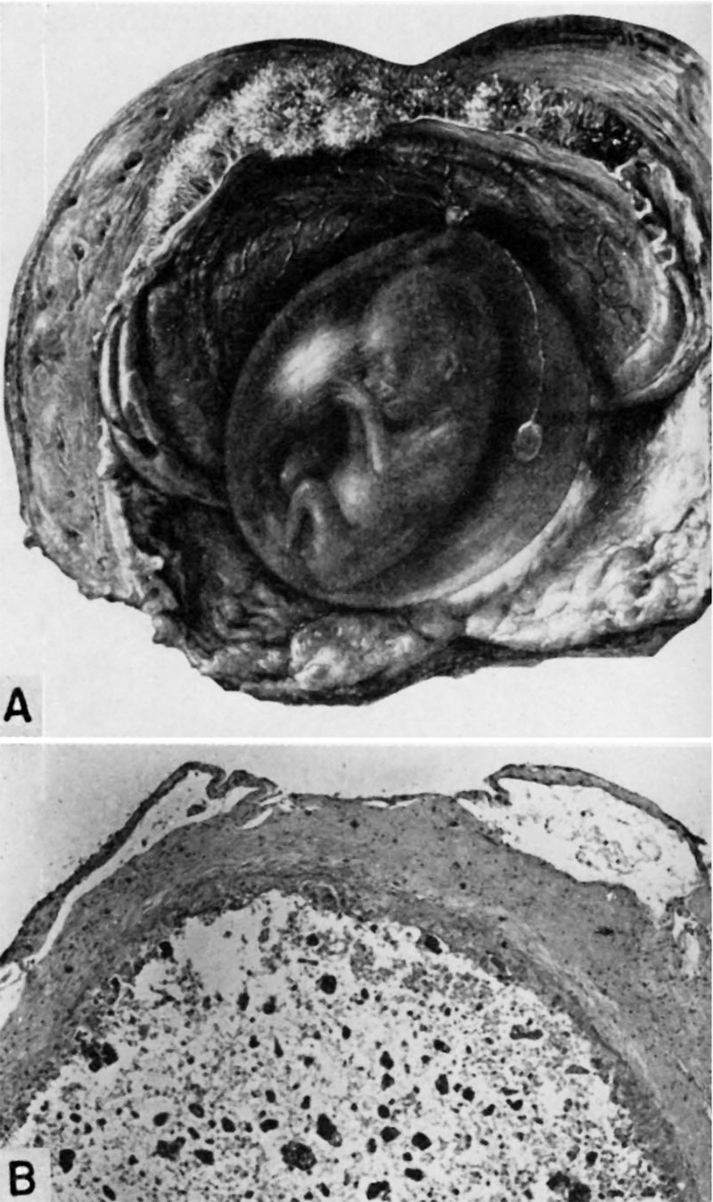
Fig. 15. Human Embryo 55 mm and older 14 Week Fetus
A half-tone drawing, natural size, of a 55 mm. embryo (menstrual age 11% weeks), lying within its amniotic sac. The chorion and uterus have been opened so as to expose the innermost membrane. The degenerating yolk-sac is seen at the right as a vesicle attached to the base of the umbilical cord by an attenuated strand—the yolkstalk. (Fig. 31 from Cullen’s “The Umbilicus and Its Diseases,” W. B. Saunders Company.)
B. A section of a slightly older yolk-sac (menstrual age 14 weeks) to show the thick avascular fibrous tissue wall and the degenerated epithelial lining. The lumen contains granular or amorphous debris which is undergoing calcification. The over; lying membrane is the amnion. B. L-i. H., S-35649, X60.
References
Hertig AT. lnvolution of tissues in fetal life: a review. (1946) Anat. Rec. 94: 96-116.
Cite this page: Hill, M.A. (2024, April 28) Embryology Hertig1946b fig15.jpg. Retrieved from https://embryology.med.unsw.edu.au/embryology/index.php/File:Hertig1946b_fig15.jpg
- © Dr Mark Hill 2024, UNSW Embryology ISBN: 978 0 7334 2609 4 - UNSW CRICOS Provider Code No. 00098G
File history
Click on a date/time to view the file as it appeared at that time.
| Date/Time | Thumbnail | Dimensions | User | Comment | |
|---|---|---|---|---|---|
| current | 08:54, 8 August 2017 | | 800 × 1,348 (196 KB) | Z8600021 (talk | contribs) |
You cannot overwrite this file.
File usage
There are no pages that use this file.