File:Hertig1946b fig14.jpg
Original file (800 × 1,273 pixels, file size: 238 KB, MIME type: image/jpeg)
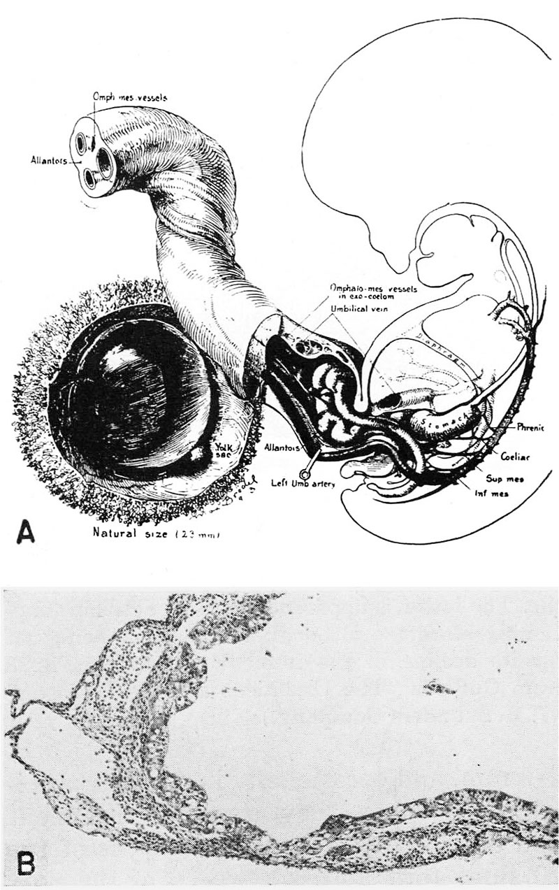
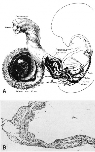
Fig. 14. Human Embryo 23 mm (week 6)
A. A drawing of a 23 mm embryo, late in the 6th week of development (menstrual age 8 weeks), showing its yolk-sac lying between the amnion and chorion. The stalk of the yolk-sac is becoming progressively more attenuated although it still contains functioning blood vessels. (Fig. 11 from Cullen’s "The Umbilicus and Its Diseases,” W. B. Saunders Company.)
B. A section of a yolk-sac whose age is comparable to that shown in fig. A. Note the thick epithelium lining the structure and the congested blood vessels within its walls. B. L-i. H., S-35-835, X 100.
References
Hertig AT. lnvolution of tissues in fetal life: a review. (1946) Anat. Rec. 94: 96-116.
Cite this page: Hill, M.A. (2024, April 27) Embryology Hertig1946b fig14.jpg. Retrieved from https://embryology.med.unsw.edu.au/embryology/index.php/File:Hertig1946b_fig14.jpg
- © Dr Mark Hill 2024, UNSW Embryology ISBN: 978 0 7334 2609 4 - UNSW CRICOS Provider Code No. 00098G
File history
Click on a date/time to view the file as it appeared at that time.
| Date/Time | Thumbnail | Dimensions | User | Comment | |
|---|---|---|---|---|---|
| current | 08:50, 8 August 2017 | | 800 × 1,273 (238 KB) | Z8600021 (talk | contribs) | ===References=== {{Ref-Hertig1946b}} {{Footer}} |
You cannot overwrite this file.
File usage
There are no pages that use this file.