File:Hertig1946b fig11.jpg: Difference between revisions
From Embryology
(===References=== {{Ref-Hertig1946b}} {{Footer}}) |
mNo edit summary |
||
| Line 1: | Line 1: | ||
==Fig. 11. A drawing of a 6.9 mm embryo in its 4th week of development== | |||
Drawn somewhat diagrammatically—to show the further approximation of the omphalo-mesenteric duct and the bodystalk due to the expanding omnion. (Fig. 4 from Cullen’s "The Umbilicus and Its Diseases,” W. B. Saunders Company.) Note that the yolk-sac is now almost as large as the amnion, the former now approaching its period of maximum activity. | |||
===References=== | ===References=== | ||
Latest revision as of 08:40, 8 August 2017
Fig. 11. A drawing of a 6.9 mm embryo in its 4th week of development
Drawn somewhat diagrammatically—to show the further approximation of the omphalo-mesenteric duct and the bodystalk due to the expanding omnion. (Fig. 4 from Cullen’s "The Umbilicus and Its Diseases,” W. B. Saunders Company.) Note that the yolk-sac is now almost as large as the amnion, the former now approaching its period of maximum activity.
References
Hertig AT. lnvolution of tissues in fetal life: a review. (1946) Anat. Rec. 94: 96-116.
Cite this page: Hill, M.A. (2024, May 17) Embryology Hertig1946b fig11.jpg. Retrieved from https://embryology.med.unsw.edu.au/embryology/index.php/File:Hertig1946b_fig11.jpg
- © Dr Mark Hill 2024, UNSW Embryology ISBN: 978 0 7334 2609 4 - UNSW CRICOS Provider Code No. 00098G
File history
Click on a date/time to view the file as it appeared at that time.
| Date/Time | Thumbnail | Dimensions | User | Comment | |
|---|---|---|---|---|---|
| current | 08:39, 8 August 2017 | 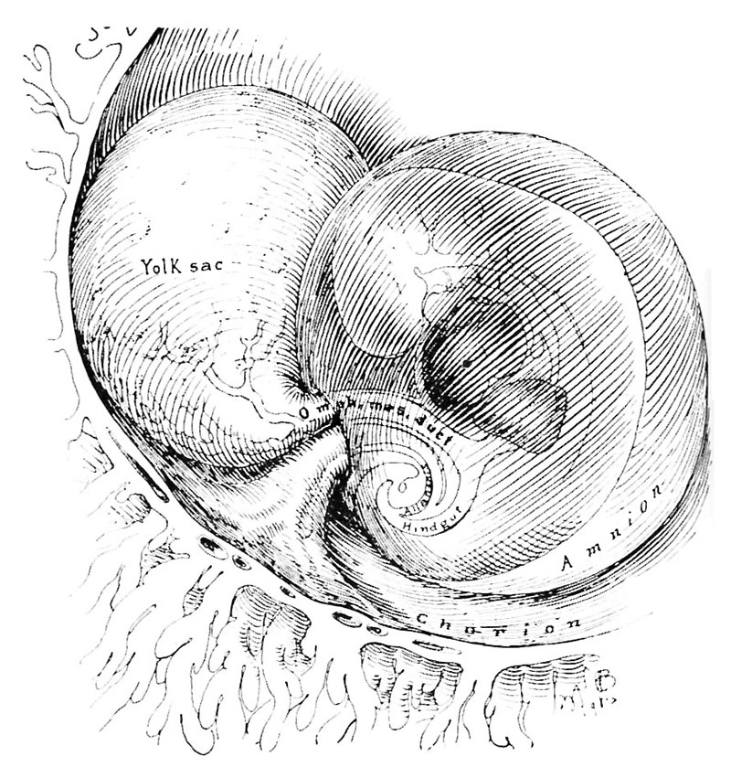 | 800 × 839 (196 KB) | Z8600021 (talk | contribs) | ===References=== {{Ref-Hertig1946b}} {{Footer}} |
You cannot overwrite this file.
File usage
The following page uses this file: