File:Hertig1946b fig10a.jpg
Hertig1946b_fig10a.jpg (800 × 515 pixels, file size: 31 KB, MIME type: image/jpeg)
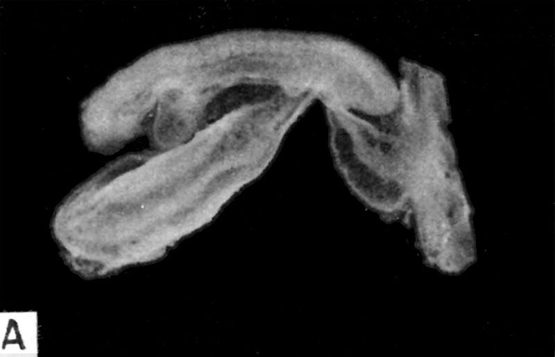
Fig. 10A. Human 3.5 mm embryo possessing 13 somites
A 3.5 mm. embryo possessing 13 somites and representing the middle part of the fourth week of development. The amnion has been removed so that the approximation of the bodystalk and omphalo-mesenteric duct are less evident than in Fig. 11. This embryo is apparently a little longer than its developmental age would allow because its natural curvature is less, perhaps from having had the amnion removed. Carnegie 6344, sequence 4, X15.
References
Hertig AT. lnvolution of tissues in fetal life: a review. (1946) Anat. Rec. 94: 96-116.
Cite this page: Hill, M.A. (2024, April 27) Embryology Hertig1946b fig10a.jpg. Retrieved from https://embryology.med.unsw.edu.au/embryology/index.php/File:Hertig1946b_fig10a.jpg
- © Dr Mark Hill 2024, UNSW Embryology ISBN: 978 0 7334 2609 4 - UNSW CRICOS Provider Code No. 00098G
File history
Click on a date/time to view the file as it appeared at that time.
| Date/Time | Thumbnail | Dimensions | User | Comment | |
|---|---|---|---|---|---|
| current | 08:32, 8 August 2017 |  | 800 × 515 (31 KB) | Z8600021 (talk | contribs) |
You cannot overwrite this file.
File usage
The following page uses this file:
