File:Hertig1946b fig03.jpg
Original file (800 × 615 pixels, file size: 132 KB, MIME type: image/jpeg)
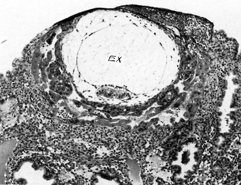
Fig. 3. A human ovum of approximately 12 days of age
The exocoelom (EX) now fills amost the entire chorionic cavity although its membranous envelope is being stretched to near the breaking point as a consequence of which it is becoming thinned out and attenuated. This stage precedes by some hours that of the 13% day ovum seen in fig. 4 at which stage the exocoelomic membrane has fragmented. Carnegie 7700, section 6-1-5, X100.
References
Hertig AT. lnvolution of tissues in fetal life: a review. (1946) Anat. Rec. 94: 96-116.
Cite this page: Hill, M.A. (2024, April 27) Embryology Hertig1946b fig03.jpg. Retrieved from https://embryology.med.unsw.edu.au/embryology/index.php/File:Hertig1946b_fig03.jpg
- © Dr Mark Hill 2024, UNSW Embryology ISBN: 978 0 7334 2609 4 - UNSW CRICOS Provider Code No. 00098G
File history
Click on a date/time to view the file as it appeared at that time.
| Date/Time | Thumbnail | Dimensions | User | Comment | |
|---|---|---|---|---|---|
| current | 16:40, 7 August 2017 | | 800 × 615 (132 KB) | Z8600021 (talk | contribs) |
You cannot overwrite this file.
File usage
The following page uses this file: