File:Hertig1946b fig02.jpg

Original file (800 × 631 pixels, file size: 153 KB, MIME type: image/jpeg)
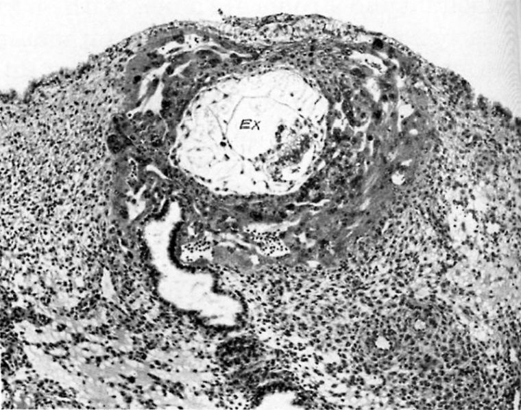
Fig. 2. A human ovum of approximately 11 days of age
The endometrial epithelium has superficially repaired the defect made by the implanting ovum. The exocoelom (EX) is now a much more definite space and is bounded below by the primitive entoderm of the bilaminar embryonic disk. Elsewhere it is surrounded by the sharply delineated exocoelomic membrane. Note the multiple gossamer attachments of the membrane by mesoblastic cells to the surrounding trophoblast from which it originally arose. Carnegie 7699, 8-5-3, X100.
References
Hertig AT. lnvolution of tissues in fetal life: a review. (1946) Anat. Rec. 94: 96-116.
Cite this page: Hill, M.A. (2024, April 27) Embryology Hertig1946b fig02.jpg. Retrieved from https://embryology.med.unsw.edu.au/embryology/index.php/File:Hertig1946b_fig02.jpg
- © Dr Mark Hill 2024, UNSW Embryology ISBN: 978 0 7334 2609 4 - UNSW CRICOS Provider Code No. 00098G
File history
Click on a date/time to view the file as it appeared at that time.
| Date/Time | Thumbnail | Dimensions | User | Comment | |
|---|---|---|---|---|---|
| current | 16:35, 7 August 2017 | | 800 × 631 (153 KB) | Z8600021 (talk | contribs) |
You cannot overwrite this file.
File usage
The following 2 pages use this file: