File:Hertig1945d fig15.jpg
From Embryology
No higher resolution available.
Hertig1945d_fig15.jpg (700 × 482 pixels, file size: 40 KB, MIME type: image/jpeg)
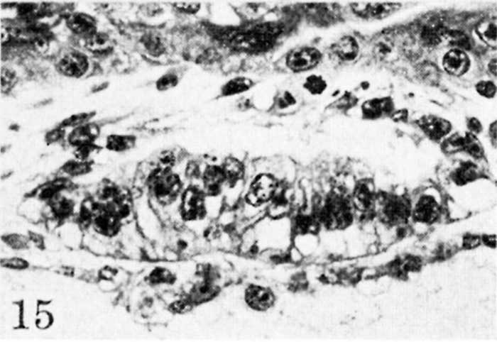
Fig. 15. Para-median section of the germ-disk and amniotic cavity from the 12.5-day ovum
Note the variable thickness and appearance of the amniogenic cells which are being added to by dividing cells derived from the adjacent trophoblast. Carnegie 7700, section S-7-7, x 250
Reference
Hertig AT. On the development of the amnion and exocoelomic membrane in the previllous human ovum. (1945) Yale J Biol Med. 18:107-15. PubMed 21007544
Cite this page: Hill, M.A. (2024, April 28) Embryology Hertig1945d fig15.jpg. Retrieved from https://embryology.med.unsw.edu.au/embryology/index.php/File:Hertig1945d_fig15.jpg
- © Dr Mark Hill 2024, UNSW Embryology ISBN: 978 0 7334 2609 4 - UNSW CRICOS Provider Code No. 00098G
File history
Click on a date/time to view the file as it appeared at that time.
| Date/Time | Thumbnail | Dimensions | User | Comment | |
|---|---|---|---|---|---|
| current | 15:48, 24 October 2017 | | 700 × 482 (40 KB) | Z8600021 (talk | contribs) |
You cannot overwrite this file.
File usage
The following 3 pages use this file: