File:Hertig1945d fig08.jpg: Difference between revisions
From Embryology
No edit summary |
mNo edit summary |
||
| (One intermediate revision by the same user not shown) | |||
| Line 1: | Line 1: | ||
==Fig. 8. A mid-cross section of an ovum of 8 to 9 days of age== | |||
The amniotic cavity is a mere slit although the amniogenic cells are well formed and have become detached from the adjacent trophoblast. The exocoelomic membrane is similar in appearance and stage of development to that of specimen shown in fig. 7. Carnegie {{CE8215}}, section 12-4-5, X 140. | |||
<gallery> | |||
File:Hertig1945d fig06.jpg|Fig 6 {{CE8155}} | |||
File:Hertig1945d fig07.jpg|Fig 7 {{CE8171}} | |||
File:Hertig1945d fig08.jpg|Fig 8 {{CE8215}} | |||
File:Hertig1945d fig09.jpg|Fig 9 {{CE8004}} | |||
</gallery> | |||
===Reference=== | |||
{{Ref-Hertig1945d}} | |||
{{Footer}} | |||
[[Category:Carnegie Embryo 8215]] | |||
Latest revision as of 15:41, 24 October 2017
Fig. 8. A mid-cross section of an ovum of 8 to 9 days of age
The amniotic cavity is a mere slit although the amniogenic cells are well formed and have become detached from the adjacent trophoblast. The exocoelomic membrane is similar in appearance and stage of development to that of specimen shown in fig. 7. Carnegie 8215, section 12-4-5, X 140.


Reference
Hertig AT. On the development of the amnion and exocoelomic membrane in the previllous human ovum. (1945) Yale J Biol Med. 18:107-15. PubMed 21007544
Cite this page: Hill, M.A. (2024, May 26) Embryology Hertig1945d fig08.jpg. Retrieved from https://embryology.med.unsw.edu.au/embryology/index.php/File:Hertig1945d_fig08.jpg
- © Dr Mark Hill 2024, UNSW Embryology ISBN: 978 0 7334 2609 4 - UNSW CRICOS Provider Code No. 00098G
File history
Click on a date/time to view the file as it appeared at that time.
| Date/Time | Thumbnail | Dimensions | User | Comment | |
|---|---|---|---|---|---|
| current | 15:38, 24 October 2017 | 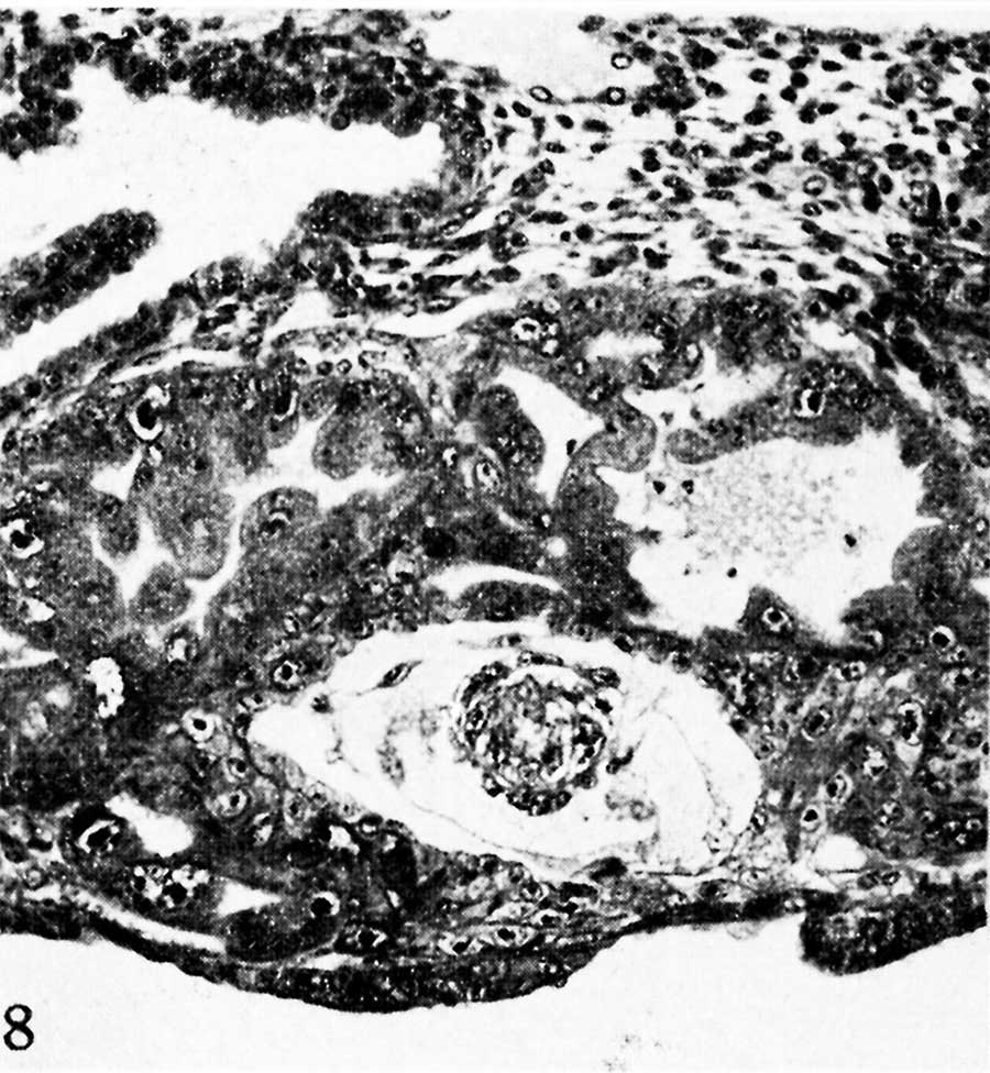 | 900 × 976 (144 KB) | Z8600021 (talk | contribs) |
You cannot overwrite this file.