File:Hassall1849 plate79.jpg

Original file (1,280 × 2,192 pixels, file size: 622 KB, MIME type: image/jpeg)
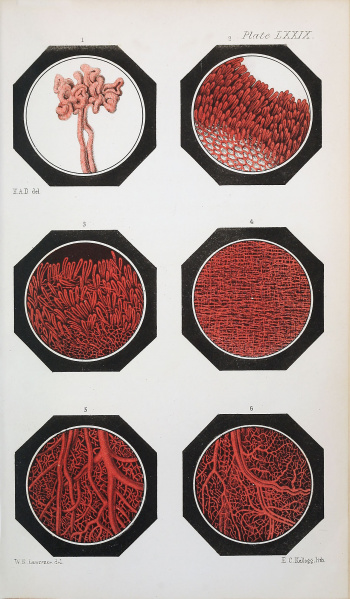
Plate LXXIX.
Fig. 1. A tuft from the fetal portion of the human placenta. 45 diameters.
Fig. 2. Papillae of the gum: a portion of the tooth is represented to exhibit the manner in which the papillae surround it. From an injection by Dr. Neill. 45 diameters.
Fig. 3. Papillae from the lip: these are observed to be rather longer and more prominent than in the gum. From an injection by Dr. Neill. 45 diameters.
Fig. 4. The arrangement of blood-vessels in the mucous membrane of the trachea. 45 diameters.
Fig. 5. shows the vascularity of the buccal membrane. 60 diameters
Fig. 6. shows the vascularity of the mucous membrane of the bladder 60 diameters.
Reference
Hassall AH. The microscopic anatomy of the human body, in health and disease. (1849) Samuel Hurley, Fleet Street, London.
Cite this page: Hill, M.A. (2024, April 27) Embryology Hassall1849 plate79.jpg. Retrieved from https://embryology.med.unsw.edu.au/embryology/index.php/File:Hassall1849_plate79.jpg
- © Dr Mark Hill 2024, UNSW Embryology ISBN: 978 0 7334 2609 4 - UNSW CRICOS Provider Code No. 00098G
File history
Click on a date/time to view the file as it appeared at that time.
| Date/Time | Thumbnail | Dimensions | User | Comment | |
|---|---|---|---|---|---|
| current | 09:23, 25 January 2019 | | 1,280 × 2,192 (622 KB) | Z8600021 (talk | contribs) | |
| 09:22, 25 January 2019 |  | 2,078 × 3,559 (1.08 MB) | Z8600021 (talk | contribs) | ||
| 09:18, 25 January 2019 |  | 2,078 × 3,559 (1.14 MB) | Z8600021 (talk | contribs) |
You cannot overwrite this file.
File usage
The following page uses this file: