File:Hassall1849 plate41 fig10.jpg
Original file (364 × 1,000 pixels, file size: 73 KB, MIME type: image/jpeg)
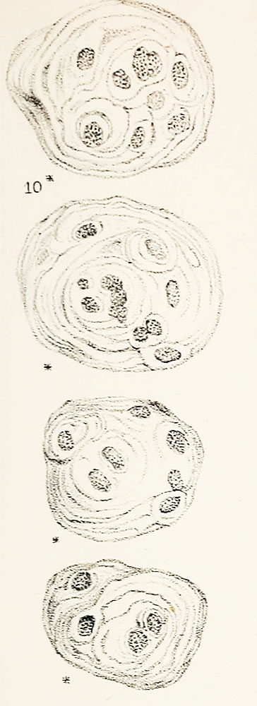
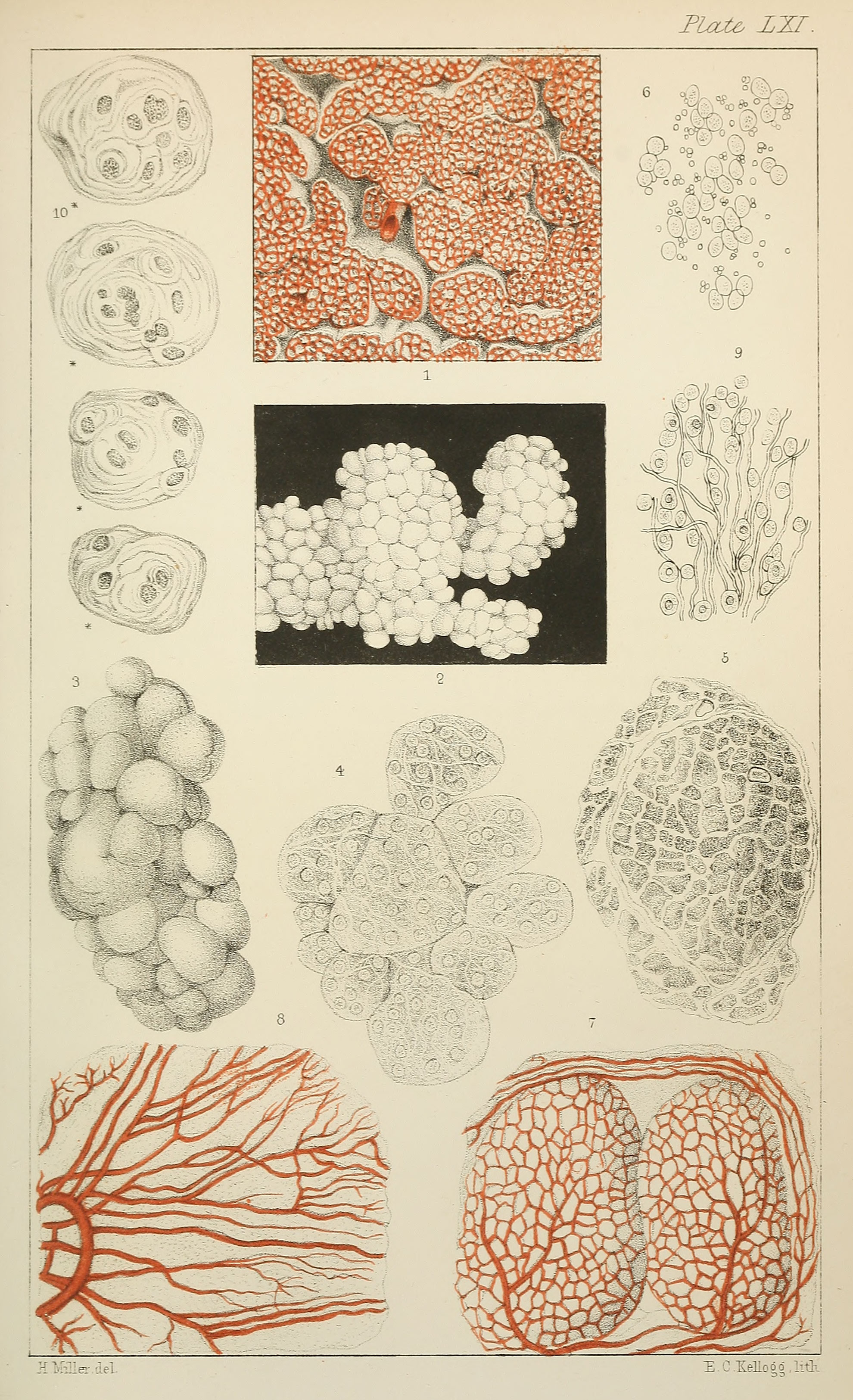
Plate LXI. Fig. 10. Compound cells of thymus
Magnified 378 diameters.
original text source for identification of "Hassall's corpuscles" within the thymus.
- "The "milky fluid" contained in the follicles and reservoir is made up, to a great extent, of an immense number of granular nuclei, as well as numerous cells of large size, which do not appear hitherto to have been either described or figured in a satisfactory manner, and which are probably to be regarded in the light of parent cells. (See Plate LXI. fig. 10.)
- Many of these cells contain several granular nuclei, each of which is surrounded by one or more concentric lamellae ; they thus resemble the cartilage cells found in the inter-vertebral substance, and also certain species of Microcystis, a genus of Fresh-water Algae."
Online Editor - This historic textbook figure is often cited as the original source for "Hassall's corpuscles" within the thymus. Note the reticular cells form an "onion-skin" arrangement of cytoplasmic layers.
Original scanned file name - Microscopicanato12hass_orig_0811.jp2
Reference
Hassall AH. The microscopic anatomy of the human body, in health and disease. (1849) Samuel Hurley, Fleet Street, London.
Cite this page: Hill, M.A. (2024, May 20) Embryology Hassall1849 plate41 fig10.jpg. Retrieved from https://embryology.med.unsw.edu.au/embryology/index.php/File:Hassall1849_plate41_fig10.jpg
- © Dr Mark Hill 2024, UNSW Embryology ISBN: 978 0 7334 2609 4 - UNSW CRICOS Provider Code No. 00098G
File history
Click on a date/time to view the file as it appeared at that time.
| Date/Time | Thumbnail | Dimensions | User | Comment | |
|---|---|---|---|---|---|
| current | 11:41, 22 January 2019 | 364 × 1,000 (73 KB) | Z8600021 (talk | contribs) | ||
| 11:37, 22 January 2019 | 500 × 1,375 (101 KB) | Z8600021 (talk | contribs) |
You cannot overwrite this file.
File usage
The following 2 pages use this file: