File:Hassall1849 plate41 fig10.jpg
Original file (364 × 1,000 pixels, file size: 73 KB, MIME type: image/jpeg)
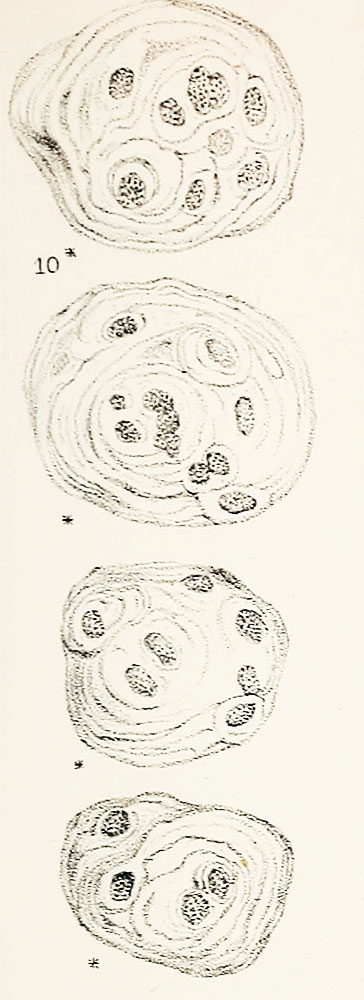
Plate LXI. Fig. 10. Compound cells of thymus
Magnified 378 diameters.
- "The "milky fluid" contained in the follicles and reservoir is made
up, to a great extent, of an immense number of granular nuclei, as well as numerous cells of large size, which do not appear hitherto to have been either described or figured in a satisfactory manner, and which are probably to be regarded in the light of parent cells. (See Plate LXI. fig. 10.)
- Many of these cells contain several granular nuclei, each of which
is surrounded by one or more concentric lamellae ; they thus resemble the cartilage cells found in the inter- vertebral substance, and also certain species of Microcystis, a genus of Fresh- water Algae."
Online Editor - This historic textbook figure is often cited as the original source for "Hassall's corpuscles" within the thymus. original text source for identification of "Hassall's corpuscles" within the thymus. Note the reticular cells form an "onion-skin" arrangement of cytoplasmic layers.
Original scanned file name - Microscopicanato12hass_orig_0811.jp2
Reference
Hassall AH. The microscopic anatomy of the human body, in health and disease. (1849) Samuel Hurley, Fleet Street, London.
Cite this page: Hill, M.A. (2024, May 20) Embryology Hassall1849 plate41 fig10.jpg. Retrieved from https://embryology.med.unsw.edu.au/embryology/index.php/File:Hassall1849_plate41_fig10.jpg
- © Dr Mark Hill 2024, UNSW Embryology ISBN: 978 0 7334 2609 4 - UNSW CRICOS Provider Code No. 00098G
File history
Click on a date/time to view the file as it appeared at that time.
| Date/Time | Thumbnail | Dimensions | User | Comment | |
|---|---|---|---|---|---|
| current | 11:41, 22 January 2019 | 364 × 1,000 (73 KB) | Z8600021 (talk | contribs) | ||
| 11:37, 22 January 2019 | 500 × 1,375 (101 KB) | Z8600021 (talk | contribs) |
You cannot overwrite this file.
File usage
The following 2 pages use this file: