File:HansonAnson1962 fig05.jpg
Original file (1,280 × 604 pixels, file size: 225 KB, MIME type: image/jpeg)
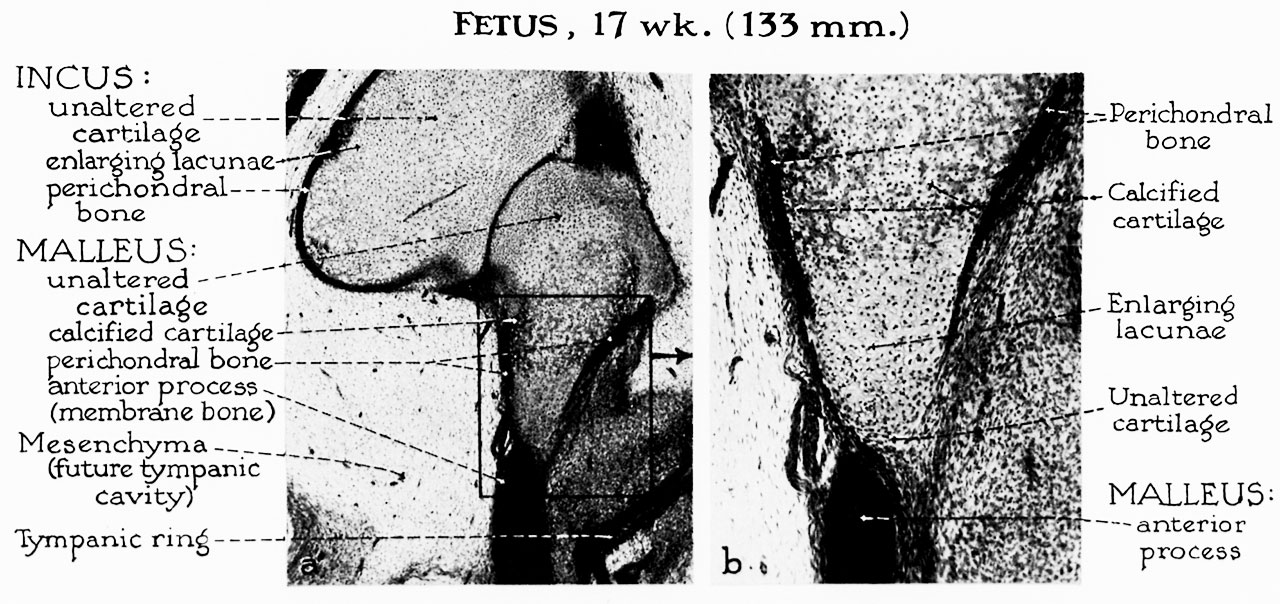
Fig. 5. Fetus 17 week 133 mm
a, By the 17-week stage, perichondral ossification has spread to the lateral aspect of the malleus, accompanied by widespread enlargement of cartilage cells and early calcification of the matrix. Endochondral bone-formation follows beneath the perichondral shell. Exceptions will occur at the articular surface, in areas of ligamentous attachment and at the distal extremity of the manubrium. Each of these will retain a thin, cartilaginous layer rather than a periosteal shell.
b, The area shown at higher magnification reveals that the anterior process of the malleus is formed in membrane bone. The process is in close proximity to the neck of the malleus but separated therefrom by fibrous tissue. Fusion will take place when ossification has been completed in the adjacent portion of the malleus.
Reference
Hanson JR. and Anson BJ. Development of the malleus of the human ear; Illustrated in atlas series. (1962) Q Bull Northwest Univ Med Sch. 36(2): 119–137. PMID: 13904457.
Cite this page: Hill, M.A. (2024, April 27) Embryology HansonAnson1962 fig05.jpg. Retrieved from https://embryology.med.unsw.edu.au/embryology/index.php/File:HansonAnson1962_fig05.jpg
- © Dr Mark Hill 2024, UNSW Embryology ISBN: 978 0 7334 2609 4 - UNSW CRICOS Provider Code No. 00098G
File history
Click on a date/time to view the file as it appeared at that time.
| Date/Time | Thumbnail | Dimensions | User | Comment | |
|---|---|---|---|---|---|
| current | 10:31, 7 January 2019 | | 1,280 × 604 (225 KB) | Z8600021 (talk | contribs) | |
| 10:28, 7 January 2019 | 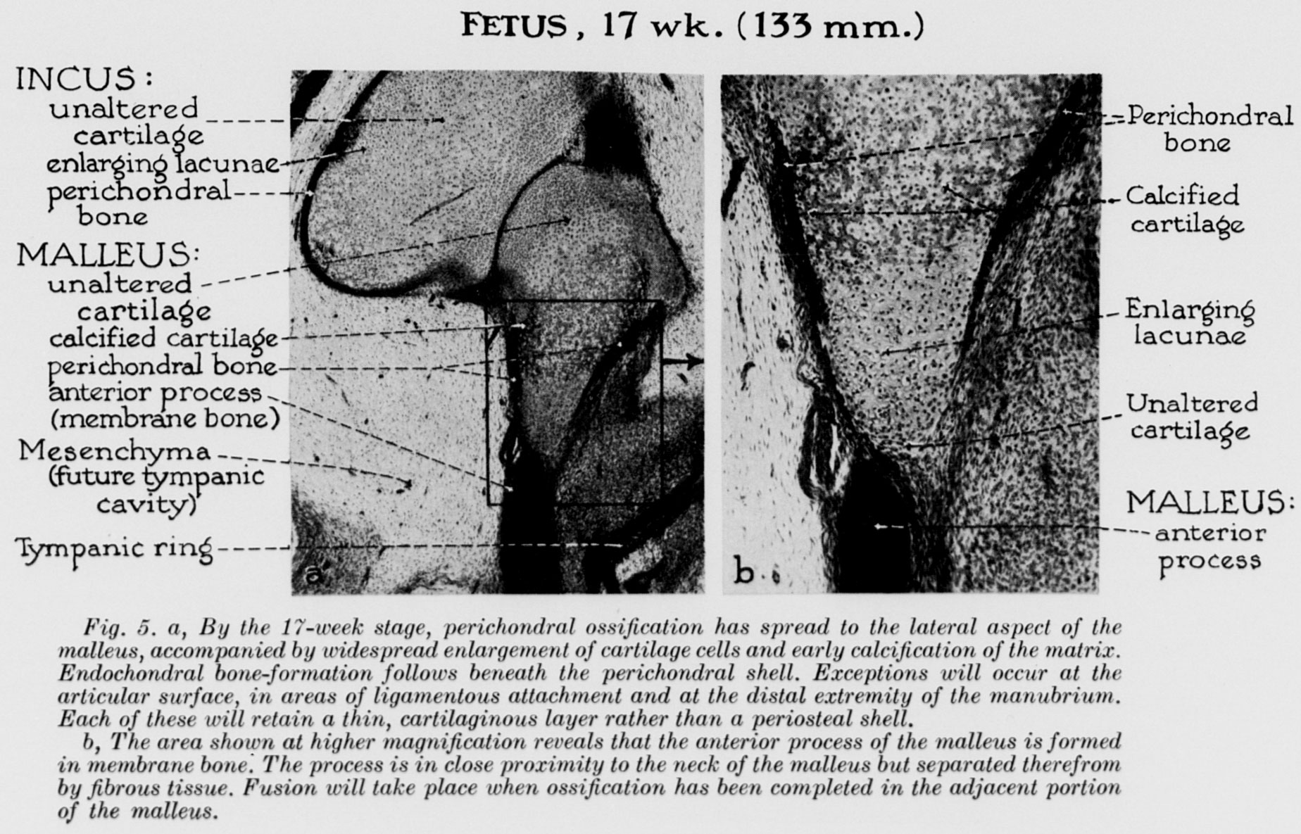 | 1,848 × 1,185 (428 KB) | Z8600021 (talk | contribs) |
You cannot overwrite this file.
File usage
The following 2 pages use this file: