File:Hamster uterus GDF8 expression.jpg
Hamster_uterus_GDF8_expression.jpg (800 × 423 pixels, file size: 57 KB, MIME type: image/jpeg)
Hamster uterus Growth-differentiation factor 8 (GDF8) Expression
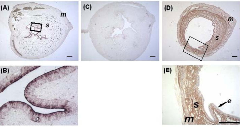
Localization of GDF-8 transcripts by in situ hybridization (A and B) and peptide by immunohistochemistry (C and D) in the hamster 72 h p.c. uterus. Transforming growth factor-beta superfamily novel member, growth-differentiation factor 8 (GDF-8).
(A) Low magnification of uterine cross section to show that GDF-8 mRNA was confined to endometrial epithelium.
(B) Insert magnified to show intense signals in epithelial cells.
(C) Absence of signals in negative control of ISH with the sense probe.
(D and E) Immunostaining localized GDF-8 peptide to the endometrial stroma (s), but not the myometrium (m) and epithelium (e)
(Number of animals =3). Bar = 100 μm).
Original file name: Figure 7. 1477-7827-7-134-7-l.jpg
Reference
<pubmed>19930721</pubmed>| Reprod Biol Endocrinol.
Wong et al. Reproductive Biology and Endocrinology 2009 7:134 doi:10.1186/1477-7827-7-134
© 2009 Wong et al; licensee BioMed Central Ltd.
This is an Open Access article distributed under the terms of the Creative Commons Attribution License (http://creativecommons.org/licenses/by/2.0), which permits unrestricted use, distribution, and reproduction in any medium, provided the original work is properly cited.
File history
Click on a date/time to view the file as it appeared at that time.
| Date/Time | Thumbnail | Dimensions | User | Comment | |
|---|---|---|---|---|---|
| current | 09:11, 26 April 2011 |  | 800 × 423 (57 KB) | S8600021 (talk | contribs) | ==Hamster uterus Growth-differentiation factor 8 (GDF8) Expression== Localization of GDF-8 transcripts by in situ hybridization (A and B) and peptide by immunohistochemistry (C and D) in the hamster 72 h p.c. uterus. Transforming growth factor-beta super |
You cannot overwrite this file.
File usage
The following page uses this file:
