File:HamiltonBoyd1960 plate13.jpg: Difference between revisions
(Z8600021 uploaded a new version of File:HamiltonBoyd1960 plate13.jpg) |
m (→Plate 13) |
||
| Line 2: | Line 2: | ||
'''Fig. 37.''' Scheme to show the essential features in placental structure which are found after the 60 mm. stage. Three cotyledons, including a marginal one, are illustrated; the cotyledons are separated from each other on the maternal side by the septa. They each contain the group of villi which constitute the associated ‘foetal’ cotyledon. The villi branch freely and there are many adhesions between adjacent ones so giving a partially labyrinthine nature to the intervillous space. Such villi are shown in relief in the marginal cotyledon and in section in the adjacent one. The third cotyledon has been dissected to show the arrangement of the septa. The openings of the endometrial arteries and veins into the intervillous space through the basal plate are indicated and an attempt has been made to show the probable degree of oxygenation of the maternal blood in the intervillous space. A marginal sinus in the intervillous space has not been included in the scheme as our material does not show such a feature. | '''Fig. 37.''' Scheme to show the essential features in placental structure which are found after the 60 mm. stage. Three cotyledons, including a marginal one, are illustrated; the cotyledons are separated from each other on the maternal side by the septa. They each contain the group of villi which constitute the associated ‘foetal’ cotyledon. The villi branch freely and there are many adhesions between adjacent ones so giving a partially labyrinthine nature to the intervillous space. Such villi are shown in relief in the marginal cotyledon and in section in the adjacent one. The third cotyledon has been dissected to show the arrangement of the septa. The openings of the endometrial arteries and veins into the intervillous space through the basal plate are indicated and an attempt has been made to show the probable degree of oxygenation of the maternal blood in the intervillous space. A marginal sinus in the intervillous space has not been included in the scheme as our material does not show such a feature. | ||
{{HamiltonBoyd1960 plates}} | |||
Revision as of 12:57, 6 August 2020
Plate 13
Fig. 37. Scheme to show the essential features in placental structure which are found after the 60 mm. stage. Three cotyledons, including a marginal one, are illustrated; the cotyledons are separated from each other on the maternal side by the septa. They each contain the group of villi which constitute the associated ‘foetal’ cotyledon. The villi branch freely and there are many adhesions between adjacent ones so giving a partially labyrinthine nature to the intervillous space. Such villi are shown in relief in the marginal cotyledon and in section in the adjacent one. The third cotyledon has been dissected to show the arrangement of the septa. The openings of the endometrial arteries and veins into the intervillous space through the basal plate are indicated and an attempt has been made to show the probable degree of oxygenation of the maternal blood in the intervillous space. A marginal sinus in the intervillous space has not been included in the scheme as our material does not show such a feature.
Plates: 1 | 2 | 3 | 4 | 5 | 6 | 7 | 8 | 9 | 10 | 11 | 12 | 13
Plate 1
Plate 2
Plate 3
Plate 4
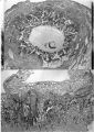
Plate 5
Plate 6
Plate 7
Plate 8
Plate 9
Plate 10
Plate 11
Plate 12

Plate 13


File history
Click on a date/time to view the file as it appeared at that time.
| Date/Time | Thumbnail | Dimensions | User | Comment | |
|---|---|---|---|---|---|
| current | 12:56, 6 August 2020 | 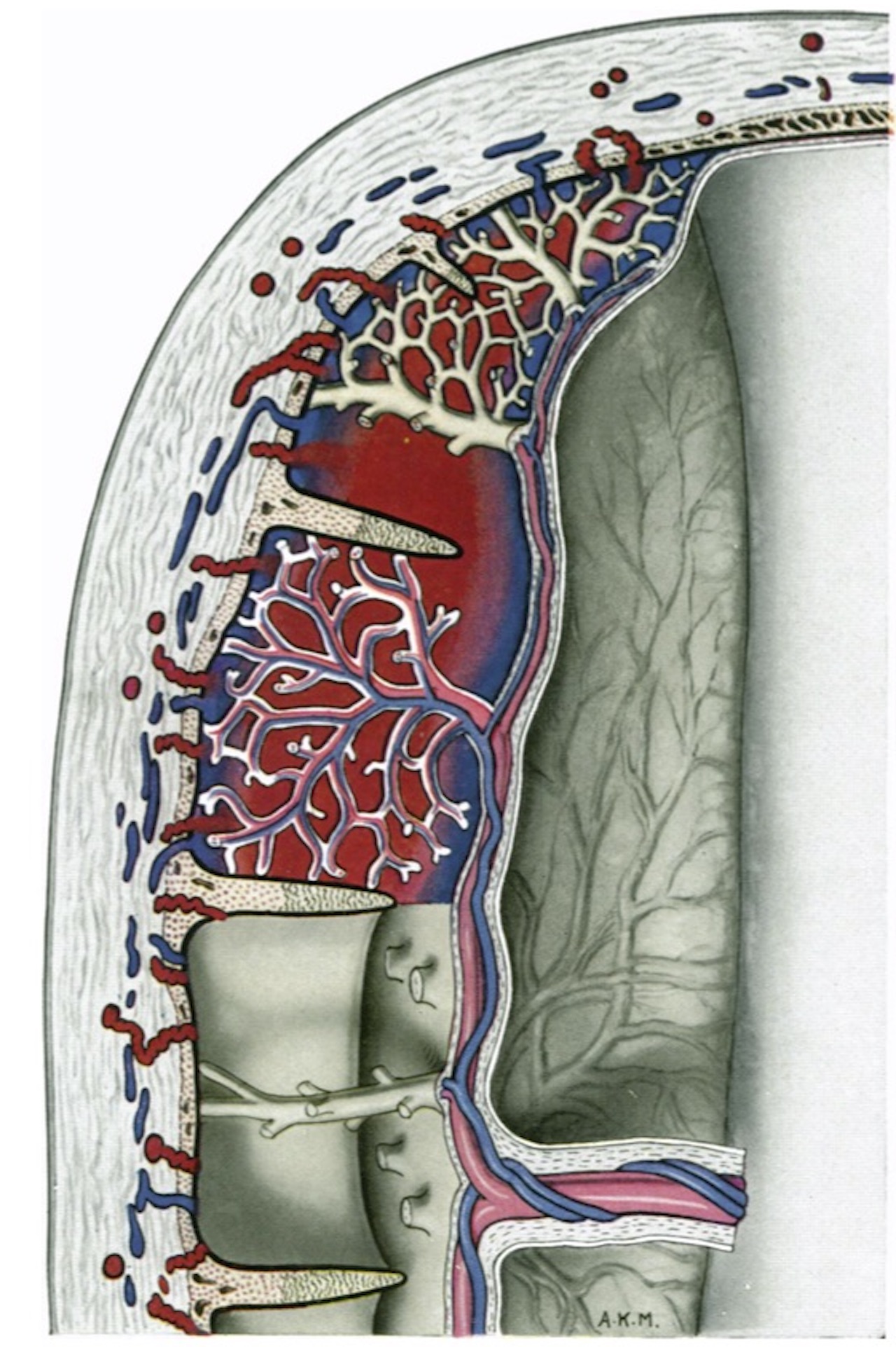 | 1,280 × 1,923 (462 KB) | Z8600021 (talk | contribs) | |
| 12:55, 6 August 2020 |  | 1,030 × 1,429 (186 KB) | Z8600021 (talk | contribs) |
You cannot overwrite this file.
File usage
The following 12 pages use this file:
- Paper - Development of the human placenta in the first three months of gestation (1960)
- File:HamiltonBoyd1960 fig02.jpg
- File:HamiltonBoyd1960 fig03.jpg
- File:HamiltonBoyd1960 fig04.jpg
- File:HamiltonBoyd1960 fig05.jpg
- File:HamiltonBoyd1960 fig06.jpg
- File:HamiltonBoyd1960 fig07.jpg
- File:HamiltonBoyd1960 fig08.jpg
- File:HamiltonBoyd1960 plate02.jpg
- File:HamiltonBoyd1960 plate03.jpg
- File:HamiltonBoyd1960 plate13.jpg
- Template:HamiltonBoyd1960 plates