File:HamiltonBoyd1960 plate12.jpg

Original file (1,280 × 1,455 pixels, file size: 605 KB, MIME type: image/jpeg)
Plate 12
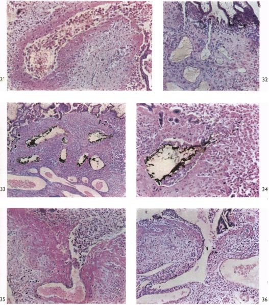
Fig. 31. Photomicrograph (x 100) of a section (Stain - Haematoxylin Eosin) of the termination of an endometrial spiral artery in the in situ placenta of a 15 mm. embryo (CX. 102). Cytotrophoblast cells are invading and plugging the lumen of the vessel. The wall of the artery shows marked degenerative changes. A portion of syncytium can be seen to the right of the illustration.
Fig. 32. Photomicrograph ( x 100) of a section (Stain - Haematoxylin Eosin) through the foetal-maternal junction in the in situ placenta of a 22 mm. embryo (CX. 103). The terminal coils of an endometrial spiral artery can be seen. A straight channel from the lumen of one of the coils extends almost to the intervillous space. Apices of villi and the cytotrophoblastic shell are seen in the upper portion of the illustration. 328 W. J. Hamilton and J. D. Boyd
Fig. 33. Photomicrograph (x 110) of a section (Stain - Haematoxylin Eosin) through the terminal coils of an endometrial spiral artery and adjacent intervillous space of a 46 mm. embryo (Camb. H. 658). Before fixation a uterine artery in this specimen was injected with indian ink. The ink can be seen in the lumen of the spiral artery and some of it is percolating amongst the cytotrophoblastic cells.
Fig. 34. Photomicrograph ( x 340) of a section through the termination of the spiral artery shown in fig. 33. The percolation of ink between the cytotrophoblastic cells is clearly seen. Note the highly modified arterial wall and the cytotrophoblastic cells within the arterial lumen.
Fig. 35. Photomicrograph ( x 70) of a section through the termination of an endometrial artery in the in situ placenta of a 50 mm. embryo (CX. 111). The arterial wall shows marked degeneration and cytotrophoblastic cells are extending into the vessel. At each side of the arterial opening Nitabuch’s membrane can be seen.
Fig. 36. Photomicrograph ( x 85) of a section of the central region of an in situ placenta of a 50 mm. embryo (CX. 111) showing communication between the intervillous space and a maternal vein. Note the continuity between cytotrophoblastic cells and unaltered venous endothelium. There is no cytotrophoblast within the lumen of the vein but a sectioned syncytial sprout can be seen.
Plates: 1 | 2 | 3 | 4 | 5 | 6 | 7 | 8 | 9 | 10 | 11 | 12 | 13
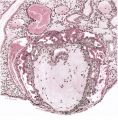
Plate 1
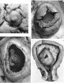
Plate 2
Plate 3
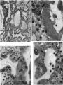
Plate 4
Plate 5
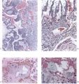
Plate 6
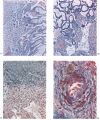
Plate 7
Plate 8
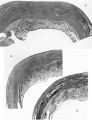
Plate 9
Plate 10
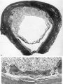
Plate 11
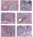
Plate 12
Plate 13


Reference
Hamilton WJ. and Boyd JD. Development of the human placenta in the first three months of gestation. (1960) J Anat. 94(3): 297-328. PMID14399291 | PDF
Cite this page: Hill, M.A. (2024, April 27) Embryology HamiltonBoyd1960 plate12.jpg. Retrieved from https://embryology.med.unsw.edu.au/embryology/index.php/File:HamiltonBoyd1960_plate12.jpg
- © Dr Mark Hill 2024, UNSW Embryology ISBN: 978 0 7334 2609 4 - UNSW CRICOS Provider Code No. 00098G
File history
Click on a date/time to view the file as it appeared at that time.
| Date/Time | Thumbnail | Dimensions | User | Comment | |
|---|---|---|---|---|---|
| current | 12:54, 6 August 2020 | | 1,280 × 1,455 (605 KB) | Z8600021 (talk | contribs) | |
| 12:52, 6 August 2020 | 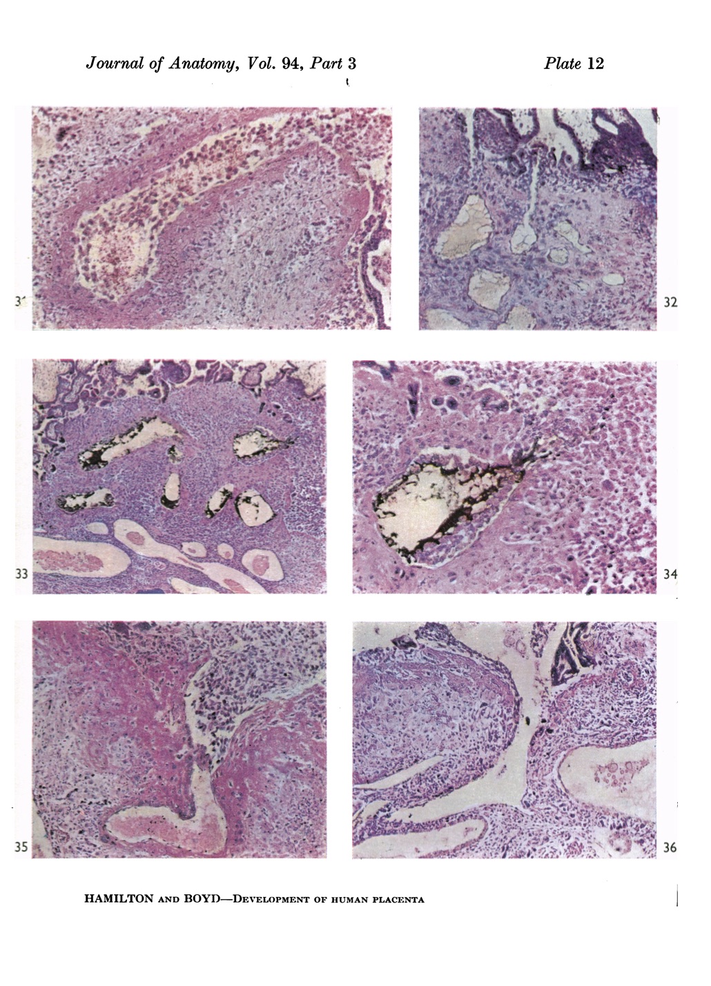 | 1,030 × 1,429 (453 KB) | Z8600021 (talk | contribs) |
You cannot overwrite this file.
File usage
The following 12 pages use this file:
- Paper - Development of the human placenta in the first three months of gestation (1960)
- File:HamiltonBoyd1960 fig02.jpg
- File:HamiltonBoyd1960 fig03.jpg
- File:HamiltonBoyd1960 fig04.jpg
- File:HamiltonBoyd1960 fig05.jpg
- File:HamiltonBoyd1960 fig06.jpg
- File:HamiltonBoyd1960 fig07.jpg
- File:HamiltonBoyd1960 fig08.jpg
- File:HamiltonBoyd1960 plate02.jpg
- File:HamiltonBoyd1960 plate03.jpg
- File:HamiltonBoyd1960 plate13.jpg
- Template:HamiltonBoyd1960 plates