File:HamiltonBoyd1960 plate10.jpg
From Embryology
Size of this preview: 331 × 599 pixels. Other resolution: 1,280 × 2,316 pixels.
Original file (1,280 × 2,316 pixels, file size: 499 KB, MIME type: image/jpeg)
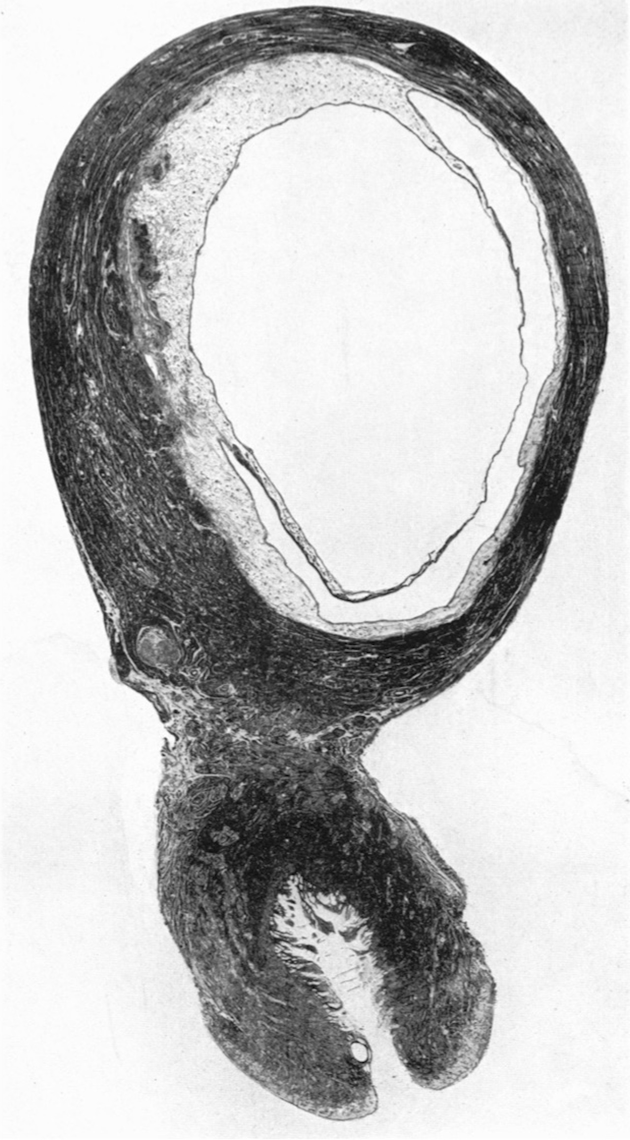
Plate 10
Fig. 28. Photograph ( x 1-8) of a uterus which contained a 30 mm. embryo (CX. 106). The decidua capsularis, which is very thin, approaches but does not make contact with the decidua vera. Note the complete absence of any indication of a marginal sinus in the intervillous space.
File history
Click on a date/time to view the file as it appeared at that time.
| Date/Time | Thumbnail | Dimensions | User | Comment | |
|---|---|---|---|---|---|
| current | 12:50, 6 August 2020 | | 1,280 × 2,316 (499 KB) | Z8600021 (talk | contribs) | |
| 12:48, 6 August 2020 |  | 1,030 × 1,429 (218 KB) | Z8600021 (talk | contribs) |
You cannot overwrite this file.
File usage
The following 12 pages use this file:
- Paper - Development of the human placenta in the first three months of gestation (1960)
- File:HamiltonBoyd1960 fig02.jpg
- File:HamiltonBoyd1960 fig03.jpg
- File:HamiltonBoyd1960 fig04.jpg
- File:HamiltonBoyd1960 fig05.jpg
- File:HamiltonBoyd1960 fig06.jpg
- File:HamiltonBoyd1960 fig07.jpg
- File:HamiltonBoyd1960 fig08.jpg
- File:HamiltonBoyd1960 plate02.jpg
- File:HamiltonBoyd1960 plate03.jpg
- File:HamiltonBoyd1960 plate13.jpg
- Template:HamiltonBoyd1960 plates