File:HamiltonBoyd1960 fig03.jpg: Difference between revisions
No edit summary |
mNo edit summary |
||
| Line 1: | Line 1: | ||
==Fig. 3. Embryo (Camb. H. 710) implantation site== | |||
Photograph ( x 3-5) of the surface view of the partially opened implantation site from which a 28-somite embryo (Camb. H. 710) had been removed. Chorionic villi are seen on the everted cut surface of the decidua capsularis. | |||
Photomicrographs of sections through this implantation site are illustrated in [[:File:HamiltonBoyd1960 plate05.jpg|Pl. 5]], fig. 14 and [[:File:HamiltonBoyd1960 plate07.jpg|PI. 7]], figs. 20-22. | |||
{{HamiltonBoyd1960 plates footer}} | |||
Latest revision as of 13:15, 6 August 2020
Fig. 3. Embryo (Camb. H. 710) implantation site
Photograph ( x 3-5) of the surface view of the partially opened implantation site from which a 28-somite embryo (Camb. H. 710) had been removed. Chorionic villi are seen on the everted cut surface of the decidua capsularis.
Photomicrographs of sections through this implantation site are illustrated in Pl. 5, fig. 14 and PI. 7, figs. 20-22.
Plates: 1 | 2 | 3 | 4 | 5 | 6 | 7 | 8 | 9 | 10 | 11 | 12 | 13
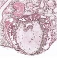
Plate 1
Plate 2
Plate 3
Plate 4
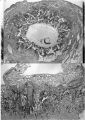
Plate 5
Plate 6
Plate 7
Plate 8
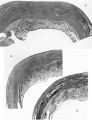
Plate 9
Plate 10
Plate 11
Plate 12
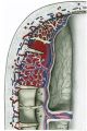
Plate 13


Reference
Hamilton WJ. and Boyd JD. Development of the human placenta in the first three months of gestation. (1960) J Anat. 94(3): 297-328. PMID14399291 | PDF
Cite this page: Hill, M.A. (2024, April 26) Embryology HamiltonBoyd1960 fig03.jpg. Retrieved from https://embryology.med.unsw.edu.au/embryology/index.php/File:HamiltonBoyd1960_fig03.jpg
- © Dr Mark Hill 2024, UNSW Embryology ISBN: 978 0 7334 2609 4 - UNSW CRICOS Provider Code No. 00098G
File history
Click on a date/time to view the file as it appeared at that time.
| Date/Time | Thumbnail | Dimensions | User | Comment | |
|---|---|---|---|---|---|
| current | 13:13, 6 August 2020 |  | 644 × 754 (114 KB) | Z8600021 (talk | contribs) |
You cannot overwrite this file.
File usage
The following 2 pages use this file: