File:Hamilton1943 fig06.jpg
From Embryology
Size of this preview: 637 × 600 pixels. Other resolution: 1,013 × 954 pixels.
Original file (1,013 × 954 pixels, file size: 110 KB, MIME type: image/jpeg)
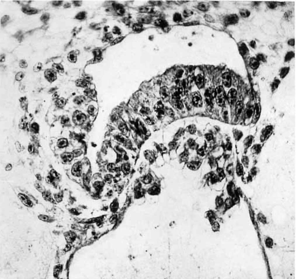
Fig. 6. A high power view of the embryonic disc
Showing the columnar nature of the embryonic ectoderm and the arrange ment of the endoderm. with Heuser’s membrane. The cells of the latter are continuous x 400.
Reference
Hamilton WJ. Barnes J. and Dodds GH. Phases of maturation, fertilization and early development in man. (1943) J. Obstet. Gynaecol, Brit. Emp., 50: 241-245.
Cite this page: Hill, M.A. (2024, May 13) Embryology Hamilton1943 fig06.jpg. Retrieved from https://embryology.med.unsw.edu.au/embryology/index.php/File:Hamilton1943_fig06.jpg
- © Dr Mark Hill 2024, UNSW Embryology ISBN: 978 0 7334 2609 4 - UNSW CRICOS Provider Code No. 00098G
File history
Click on a date/time to view the file as it appeared at that time.
| Date/Time | Thumbnail | Dimensions | User | Comment | |
|---|---|---|---|---|---|
| current | 21:02, 30 October 2017 | | 1,013 × 954 (110 KB) | Z8600021 (talk | contribs) | ==Plate 2.== Fig. 5. A surface view of the endometrium of specimen No. 3. The smooth elevation produced by the implanting embryo is seen. The endometritnn shows fissures :md crct'iccs which for the most part are associated with the mouths of the uteri... |
You cannot overwrite this file.
File usage
The following 2 pages use this file: