File:Haines1947 plate04.jpg

Original file (1,280 × 1,572 pixels, file size: 429 KB, MIME type: image/jpeg)
Plate 4
fig. 49. Hughes’s 60 mm. embryo, 7.3.8. Intercarpal joints between capitate and trapezoid. The interosseous (51.) and dorsal (d.l.) ligaments enclose between them a mass of synovial mesenchyme containing blood vessels (b.'v.).
50. Hughes’s 60 mm. embryo, 3.3.7. Inferior radio-ulnar joint. The joint cavity is spreading by liquefaction of the synovial mesenchyme (s.m.). The synovial surface is ragged, and strands of cells stretch into the cavity. Some of the cells are degenerate in appearance (d.c.).
fig. 51. 66 mm. Appleton’s ‘H 66’, 23.1. Shoulder and acromio-clavicular joint. The synovial surfaces are smooth and the humeral head is more curved than the glenoid surface. The walls of the subgacromial bursa (s.b.) have a. structure similar to those of the joint cavity. The clavicle and glenoid surface are covered with layers of articular fibro-cartilage (f.c.). 52. 66 mm. Appleton’s ‘H 66’, 24.3. Shoulder. The recess of the joint cavity showing the synovial stratum (s.s.), and the vascular sub-synovial tissue. * - ~
53. 125 mm. Appleton’s ‘H 125’, 18.1. Proximal inter-phalangeal joint of the third digit of the hand, illustrating the formation of a synovial fold (8. f.). The ragged synovial surface (r.s.) indicates that the joint cavity is still extending by liquefaction of the synovial mesenchyme.
54. 190 mm. Appleton’s ‘H 190’, 2.3. Humero-radial joint. General view showing the incongruity of the articular surfaces, the synovial folds and synovial villi. .
55. 190 mm. Appleton’s ‘ H 190’, 2.3. Same section. The articular surfaces of the cartilages show distinct fibrillar layers (f.l.), and the structure of the cartilage has not attained its adult pattern.
56. 190 mm. Appleton’s ‘H 190’, 2.3. Same section. High-power view of a synovial villus, showing also the synovial stratum (s.s.), a poorly developed sub-synovial stratum (s.b.), and a typical transition zone_ (tr.) between the articular cartilage and perichondrial tissues.
57. Hughes’s 20 day rat. The radial collateral ligament (lg.) shows twisting of its fibres and a structural similarity to the tendons of origin of the extensor muscles (t.m.).
58. Hughes’s 20 day rat. Humero-radial joint. General view showing the apparent union of the cartilages, and the structure of the fibrous capsule (cp.).
59. Hughes’s 20 day rat. Metacarpo-phalangeal joint of second digit. The cartilages are apparently joined, but in the region of union the fibrillar layer (f.l.) is conspicuous.
Reference
Haines RW. The development of joints. (1947) J. Anat. 81, 33-55.
Cite this page: Hill, M.A. (2024, April 27) Embryology Haines1947 plate04.jpg. Retrieved from https://embryology.med.unsw.edu.au/embryology/index.php/File:Haines1947_plate04.jpg
- © Dr Mark Hill 2024, UNSW Embryology ISBN: 978 0 7334 2609 4 - UNSW CRICOS Provider Code No. 00098G
File history
Click on a date/time to view the file as it appeared at that time.
| Date/Time | Thumbnail | Dimensions | User | Comment | |
|---|---|---|---|---|---|
| current | 15:00, 3 October 2017 | | 1,280 × 1,572 (429 KB) | Z8600021 (talk | contribs) | |
| 14:58, 3 October 2017 | 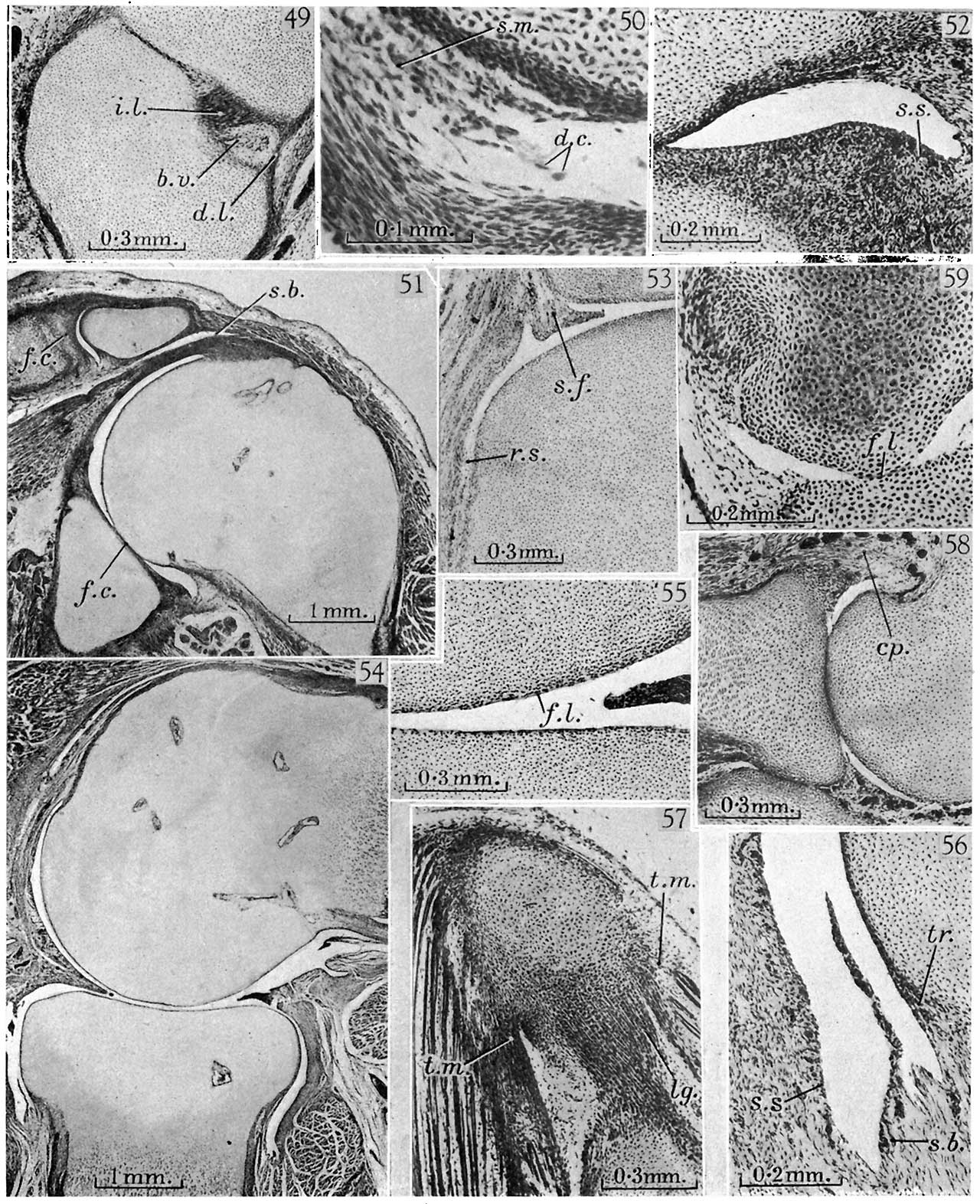 | 1,280 × 1,572 (412 KB) | Z8600021 (talk | contribs) | ||
| 14:57, 3 October 2017 |  | 1,788 × 2,422 (628 KB) | Z8600021 (talk | contribs) | {{Ref-Haines1947}} |
You cannot overwrite this file.
File usage
The following page uses this file: