File:Haines1947 plate02.jpg

Original file (1,280 × 1,586 pixels, file size: 505 KB, MIME type: image/jpeg)
Plate 2
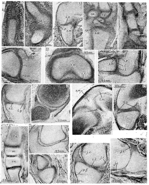
fig. 15. 16mm. Kirk’s ‘Series I’, 23.4.5. Metacarpo-phalangeal region. The metacarpal (mt.) is well chondrified and proportionately large, the basal phalanx (b. 10.) has now chondrified, but beyond this phalanx the blastema (bl.) is undiiferentiated. The joint region is surrounded by mesenchymal tissue in which the capsule has not yet appeared.
16. 16 mm. Kirk’s ‘Series I’, 41.1.6. Knee. The capsule (cp.) is well developed posteriorly and cuts off an abundant synovial mesenchyme (s.m.) with its included blood vessels (b.v.). 17. 19 mm. Appleton’s ‘H 19’, 4.3.6. Humero-radial joint. The homogeneous interzone (i.z.), intra,- and'extra-capsular perichondrium (i. p. and e.p.), synovial mesenchyme (.s.m.) and fibrous capsule (01).) take the classical form. _
18. 19 mm. Appleton’s ‘H 19’, 3.3.5. Carpus. The carpal elements, the trapezium (tm.), trapezoid (td.), scaphoid (sc.) and centrale (ce.), are separated from each other and from the radius and metacarpals by interzones of the typical form. The capsule of the wrist joint (cp.) can be distinguished enclosing the synovial mesenchyme (s.m.).
19. 19 mm. Appleton’s ‘H 19’, 3.2.3. Carpo-metacarpal and metacarpo phalangeal joints of the thumb. The fibrous capsules (cp.) are dense in these joints, for the tissue destined to form the collateral ligaments is not yet differentiated from them. The bulging curves of the capsule of the metacarpo-phalangeal joint are very clearly shown, uncomplicated by associated condensed structures.
20. 21 mm. Kirk’s ‘Series 8’, 78.1.2. Shoulder. A loose zone (l.z.) separating two dense chondrogenous layers (c.l.) can barely be distinguished in the interzone. The capsule (cp.) is now distinct and encloses a scanty synovial mesenchyme (s.m.).
21. 21 mm. Kirk’s ‘Series 8’, 90.1.3. Elbow. The three-layered interzone with its chondrogenous layers (c.l.) and loose intermediate layer (i.l.) is shown fully developed between humerus and radius and partly developed between humerus and 11lna. The chondrogenous layers are continued into the intra-capsular perichondrium (i.p.) and the intermediate layer into the synovial mesenchyme (s.m.) which lies within the capsule (cp.).
22. 21 mm. Kirk’s ‘Series 8’, 85.1.3. Knee. The condyles of the femur are separated from the tibia by homogeneous interzones (i.z.), and the intercondylar space is filled by synovial mesenchyme (s.m.). The menisci have not yet appeared. Between the femur and the quadriceps tendon is a layer of loose synovial mesenchyme (l.m.) where the joint cavity will appear at a later stage.
23. 23 mm. Appleton’s ‘H 23’, 8.2.2. Shoulder. A typical three-layered interzone has appeared at this stage. The chondrogenous layer (c.l.) covers the glenoid surface of the scapula and is continued into the labrum glenoidale (l.g.).
24. 24 mm. London Hospital ‘mx’ (Rutherford), 120.12. Shoulder. The labrum glenoidale is very distinct and is continuous with the chondrogenous layer of the interzone.
25. 26 mm. Boyd’s ‘H 11’, 75.2.1. Knee, condylar region. The interzone between the condyle of the femur and tibia is in the early three-layered stage. The menisci (mn.) are indicated by faint condensation of the mesenchyme.
26. 26 mm. Boyd’s ‘H 11’, 74.1.3. Knee, intercondylar region. In the abundant synovial mesenchyme (s.m.), which contains numerous small blood vessels, is the posterior cruciate ligament (cr.). The patella is separated from the femur by a layer of loose synovial mesenchyme (l.m.). p
27. 26 mm. Boyd’s ‘H 11 ’, 75.2.1. Tarsus. The talus (ta.), scaphoid (.90.), first cuneiform (cn.) and metatarsal are separated by homogeneous interzones.
28. 29 mm. Appleton’s ‘H 29’, 10.2.2. ‘Shoulder. Liquefaction of the synovial mesenchyme has begun at the peripheryof the joint, so that the position of the joint cavity (j.c.) is clearly defined, but the cavity is still partly filled with cells. The intermediate layer of the interzone (z'.l.) appears collapsed, and its flattened cells are clumped, but this condition is probably an artifact caused by swelling of the cartilage. A pressure fold ( p. f.) indicates swelling of the cartilage at some stage in the preparation. .
29. 29 mm. Appleton’s ‘H 29’, 26.2.1. Humero-ulnar joint. The three layers of the interzone are sharply defined and in the intermediate layer the cells have become flattened and arranged parallel to the articular surfaces. separated by a synovial cavity which still contains some scattered strands of cells (s.c.).
30. 29 mm. Appleton’s ‘H 29’, 28.2.5. Fourth metacarpo-phalangeal joint. The interzone is in the early three-layered stage. .
31. 30 mm. Appleton’s ‘H 30’, 12.2.7. Shoulder. In the peripheral parts of the joint the synovial mesenchyme is now liquefied to form the joint cavity (j.c.). More centrally the scapula and humerus appear to be in continuity, but this is probably due to an artifact, as in fig. 28.
Reference
Haines RW. The development of joints. (1947) J. Anat. 81, 33-55.
Cite this page: Hill, M.A. (2024, April 28) Embryology Haines1947 plate02.jpg. Retrieved from https://embryology.med.unsw.edu.au/embryology/index.php/File:Haines1947_plate02.jpg
- © Dr Mark Hill 2024, UNSW Embryology ISBN: 978 0 7334 2609 4 - UNSW CRICOS Provider Code No. 00098G
File history
Click on a date/time to view the file as it appeared at that time.
| Date/Time | Thumbnail | Dimensions | User | Comment | |
|---|---|---|---|---|---|
| current | 15:10, 3 October 2017 | | 1,280 × 1,586 (505 KB) | Z8600021 (talk | contribs) | |
| 15:09, 3 October 2017 |  | 1,779 × 2,413 (788 KB) | Z8600021 (talk | contribs) |
You cannot overwrite this file.
File usage
The following page uses this file: