File:Haines1947 fig03.jpg
From Embryology
No higher resolution available.
Haines1947_fig03.jpg (314 × 550 pixels, file size: 44 KB, MIME type: image/jpeg)
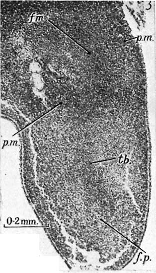
Fig. 3. 10-5 mm. Smout’s 5} weeks embryo, 16.1.4. Hind-limb
The skeletal blastema of the femoral (fm.), tibial (tb.) and foot-plate (f.p.) regions are distinguishable. The pre-muscle masses of the thigh (p.m.) are more condensed than the general mesenchyme, but those for the more distal part of the limbs have not yet appeared.
Reference
Haines RW. The development of joints. (1947) J. Anat. 81, 33-55.
Cite this page: Hill, M.A. (2024, May 21) Embryology Haines1947 fig03.jpg. Retrieved from https://embryology.med.unsw.edu.au/embryology/index.php/File:Haines1947_fig03.jpg
- © Dr Mark Hill 2024, UNSW Embryology ISBN: 978 0 7334 2609 4 - UNSW CRICOS Provider Code No. 00098G
File history
Click on a date/time to view the file as it appeared at that time.
| Date/Time | Thumbnail | Dimensions | User | Comment | |
|---|---|---|---|---|---|
| current | 15:21, 3 October 2017 | | 314 × 550 (44 KB) | Z8600021 (talk | contribs) |
You cannot overwrite this file.
File usage
There are no pages that use this file.