File:Gray1235.jpg
Gray1235.jpg (800 × 389 pixels, file size: 41 KB, MIME type: image/jpeg)
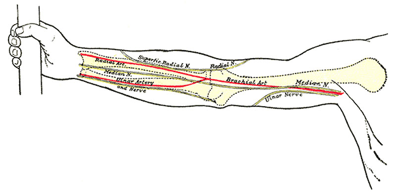
Right Upper Limb
Front of right upper extremity, showing surface markings for bones, arteries, and nerves.
Arteries
The course of the axillary artery can be marked out by abducting the arm to a right angle and drawing a line from the middle of the clavicle to the point where the tendon of the Pectoralis major crosses the prominence of the Coracobrachialis. Of the branches of the axillary artery, the origin of the thoracoacromial corresponds to the point where the artery crosses the upper border of Pectoralis minor; the lateral thoracic takes practically the line of the lower border of Pectoralis minor; the subscapular is sufficiently indicated by the axillary border of the scapula; the scapular circumflex is given off the subscapular opposite the midpoint of a line joining the tip of the acromion to the lower edge of the deltoid tuberosity, while the humeral circumflex arteries arise from the axillary about 2 cm. above this.
The position of the brachial artery is marked by a line drawn from the junction of the anterior and middle thirds of the distance between the anterior and posterior axillary folds to a point midway between the epicondyles of the humerus and continued distally for 2.5 cm., at which point the artery bifurcates. With regard to the branches of the brachial artery—the profunda crosses the back of the humerus at the level of the insertion of Deltoideus; the nutrient is given off opposite the middle of the body of the humerus; a line from this point to the back of the medial condyle represents the superior ulnar collateral; the inferior ulnar collateral is given off about 5 cm. above the fold of the elbow-joint and runs directly medialward.
Nerves
In the arm the line of the median nerve is practically the same as that for the brachial artery; at the bend of the elbow the nerve is medial to the artery. The course of the nerve in the forearm is marked by a line starting from a point just medial to the center of one joining the epicondyles, and extending to the lateral margin of the tendon of Palmaris longus at the wrist.
- Gray's Images: Development | Lymphatic | Neural | Vision | Hearing | Somatosensory | Integumentary | Respiratory | Gastrointestinal | Urogenital | Endocrine | Surface Anatomy | iBook | Historic Disclaimer
| Historic Disclaimer - information about historic embryology pages |
|---|
 |
| iBook - Gray's Embryology | |
|---|---|

|
|
Reference
Gray H. Anatomy of the human body. (1918) Philadelphia: Lea & Febiger.
Cite this page: Hill, M.A. (2024, April 27) Embryology Gray1235.jpg. Retrieved from https://embryology.med.unsw.edu.au/embryology/index.php/File:Gray1235.jpg
- © Dr Mark Hill 2024, UNSW Embryology ISBN: 978 0 7334 2609 4 - UNSW CRICOS Provider Code No. 00098G
File history
Click on a date/time to view the file as it appeared at that time.
| Date/Time | Thumbnail | Dimensions | User | Comment | |
|---|---|---|---|---|---|
| current | 11:52, 25 February 2013 | | 800 × 389 (41 KB) | Z8600021 (talk | contribs) | {{Gray Anatomy}} Category:Human Category:Limb Category:Bone |
You cannot overwrite this file.
File usage
The following page uses this file: