File:Gray1166.jpg

Original file (750 × 750 pixels, file size: 194 KB, MIME type: image/jpeg)
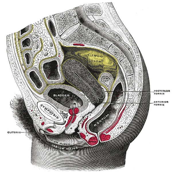
Fig. 1166
Sagittal section of the lower part of a female trunk, right segment. SM. INT. Small intestine. (Testut.)
The uterus (Figs. 1161, 1165, 1166) is a hollow, thick-walled, muscular organ situated deeply in the pelvic cavity between the bladder and rectum. Into its upper part the uterine tubes open, one on either side, while below, its cavity communicates with that of the vagina. When the ova are discharged from the ovaries they are carried to the uterine cavity through the uterine tubes. If an ovum be fertilized it imbeds itself in the uterine wall and is normally retained in the uterus until prenatal development is completed, the uterus undergoing changes in size and structure to accommodate itself to the needs of the growing embryo.
After parturition the uterus returns almost to its former condition, but certain traces of its enlargement remains. It is necessary, therefore, to describe as the type-form the adult virgin uterus, and then to consider the modifications which are effected as a result of pregnancy.
- Links: Uterus Development | Ovary Development
- Gray's Images: Development | Lymphatic | Neural | Vision | Hearing | Somatosensory | Integumentary | Respiratory | Gastrointestinal | Urogenital | Endocrine | Surface Anatomy | iBook | Historic Disclaimer
| Historic Disclaimer - information about historic embryology pages |
|---|
| Pages where the terms "Historic" (textbooks, papers, people, recommendations) appear on this site, and sections within pages where this disclaimer appears, indicate that the content and scientific understanding are specific to the time of publication. This means that while some scientific descriptions are still accurate, the terminology and interpretation of the developmental mechanisms reflect the understanding at the time of original publication and those of the preceding periods, these terms, interpretations and recommendations may not reflect our current scientific understanding. (More? Embryology History | Historic Embryology Papers) |
| iBook - Gray's Embryology | |
|---|---|

|
|
Reference
Gray H. Anatomy of the human body. (1918) Philadelphia: Lea & Febiger.
Cite this page: Hill, M.A. (2024, April 27) Embryology Gray1166.jpg. Retrieved from https://embryology.med.unsw.edu.au/embryology/index.php/File:Gray1166.jpg
- © Dr Mark Hill 2024, UNSW Embryology ISBN: 978 0 7334 2609 4 - UNSW CRICOS Provider Code No. 00098G
File history
Click on a date/time to view the file as it appeared at that time.
| Date/Time | Thumbnail | Dimensions | User | Comment | |
|---|---|---|---|---|---|
| current | 15:44, 9 June 2013 |  | 750 × 750 (194 KB) | Z8600021 (talk | contribs) | :'''Links:''' Uterus Development | Ovary Development {{Gray Anatomy}} Category:Female Category:Uterus Category:Renal |
You cannot overwrite this file.
File usage
The following page uses this file:
