File:Gray1132.jpg
Gray1132.jpg (625 × 500 pixels, file size: 86 KB, MIME type: image/jpeg)
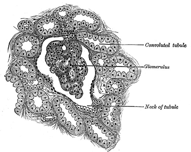
Section of cortex of human kidney
Structure of the Renal Tubules
The renal tubules consist of a basement membrane lined with epithelium. The epithelium varies considerably in different sections of the tubule. In the neck the epithelium is continuous with that lining the glomerular capsule, and like it consists of flattened cells each containing an oval nucleus (Fig. 1132). The two convoluted tubules, the spiral and zigzag tubules and the ascending limb of Henle’s loop, are lined by a type of epithelium which is histologically the same in all. The cells are somewhat columnar in shape and dovetail into one another of their lateral aspect. Each has a striated border next the lumen of the tube, its inner part is granular and its outer portion vertically striated. The nucleus is spherical and situated about the center of the cell. In the descending limb of Henle’s loop the epithelium resembles that found in the glomerular capsule and the commencement of the tube, consisting of flat, clear epithelial plates, each with an oval nucleus (Fig. 1131). The nuclei alternate on opposite surfaces of the tubule so that the lumen remains fairly constant.
In the straight tube the epithelium is clear and cubical: in its papillary portion the cells are distinctly columnar and transparent (Fig. 1132).
- Links: Renal System Development
- Gray's Images: Development | Lymphatic | Neural | Vision | Hearing | Somatosensory | Integumentary | Respiratory | Gastrointestinal | Urogenital | Endocrine | Surface Anatomy | iBook | Historic Disclaimer
| Historic Disclaimer - information about historic embryology pages |
|---|
 |
| iBook - Gray's Embryology | |
|---|---|

|
|
Reference
Gray H. Anatomy of the human body. (1918) Philadelphia: Lea & Febiger.
Cite this page: Hill, M.A. (2024, April 27) Embryology Gray1132.jpg. Retrieved from https://embryology.med.unsw.edu.au/embryology/index.php/File:Gray1132.jpg
- © Dr Mark Hill 2024, UNSW Embryology ISBN: 978 0 7334 2609 4 - UNSW CRICOS Provider Code No. 00098G
File history
Click on a date/time to view the file as it appeared at that time.
| Date/Time | Thumbnail | Dimensions | User | Comment | |
|---|---|---|---|---|---|
| current | 23:02, 17 September 2012 | | 625 × 500 (86 KB) | Z8600021 (talk | contribs) | :'''Links:''' Renal System Development {{Template:Gray Anatomy}} Category:Renal Category:Cardiovascular |
You cannot overwrite this file.
File usage
The following page uses this file: