File:Gray1120.jpg
Original file (683 × 900 pixels, file size: 297 KB, MIME type: image/jpeg)
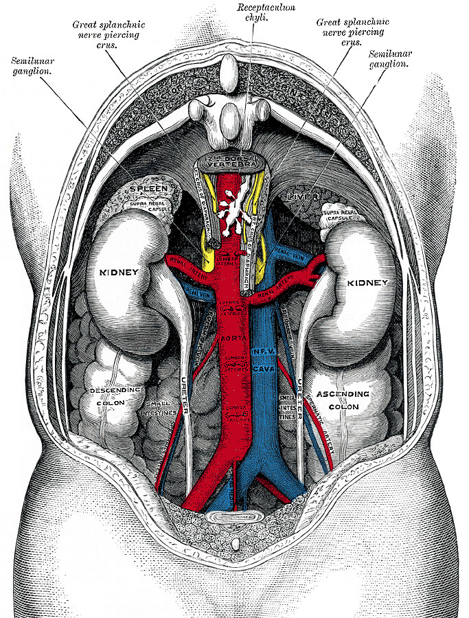
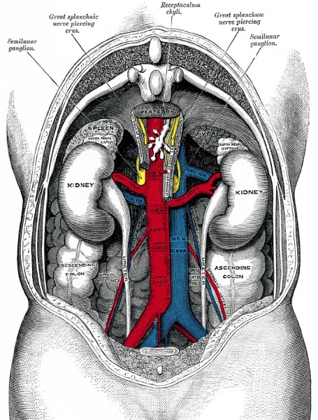
Fig. 1120 The relations of the viscera and large vessels of the abdomen
Seen from behind, the last thoracic vertebra being well raised.
Relations
The anterior surface (facies anterior) (Figs. 1120 and 1122) of each kidney is convex, and looks forward and lateralward. Its relations to adjacent viscera differ so completely on the two sides that separate descriptions are necessary.
The Posterior Surface
(facies posterior) (Figs. 1123, 1124).—The posterior surface of each kidney is directed backward and medialward. It is imbedded in areolar and fatty tissue and entirely devoid of peritoneal covering. It lies upon the diaphragm, the medial and lateral lumbocostal arches, the Psoas major, the Quadratus lumborum, and the tendon of the Transversus abdominis, the subcostal, and one or two of the upper lumbar arteries, and the last thoracic, iliohypogastric, and ilioinguinal nerves. The right kidney rests upon the twelfth rib, the left usually on the eleventh and twelfth. The diaphragm separates the kidney from the pleura, which dips down to form the phrenicocostal sinus, but frequently the muscular fibers of the diaphragm are defective or absent over a triangular area immediately above the lateral lumbocostal arch, and when this is the case the perinephric areolar tissue is in contact with the diaphragmatic pleura.
- Links: Renal System Development
- Gray's Images: Development | Lymphatic | Neural | Vision | Hearing | Somatosensory | Integumentary | Respiratory | Gastrointestinal | Urogenital | Endocrine | Surface Anatomy | iBook | Historic Disclaimer
| Historic Disclaimer - information about historic embryology pages |
|---|
 |
| iBook - Gray's Embryology | |
|---|---|

|
|
Reference
Gray H. Anatomy of the human body. (1918) Philadelphia: Lea & Febiger.
Cite this page: Hill, M.A. (2024, April 27) Embryology Gray1120.jpg. Retrieved from https://embryology.med.unsw.edu.au/embryology/index.php/File:Gray1120.jpg
- © Dr Mark Hill 2024, UNSW Embryology ISBN: 978 0 7334 2609 4 - UNSW CRICOS Provider Code No. 00098G
File history
Click on a date/time to view the file as it appeared at that time.
| Date/Time | Thumbnail | Dimensions | User | Comment | |
|---|---|---|---|---|---|
| current | 16:51, 16 September 2012 | | 683 × 900 (297 KB) | Z8600021 (talk | contribs) | ==The relations of the viscera and large vessels of the abdomen== Seen from behind, the last thoracic vertebra being well raised. {{Template:Gray Anatomy}} Category:Renal Category:Cardiovascular |
You cannot overwrite this file.
File usage
The following page uses this file: