File:Gray1115.jpg
From Embryology
Gray1115.jpg (600 × 474 pixels, file size: 73 KB, MIME type: image/jpeg)
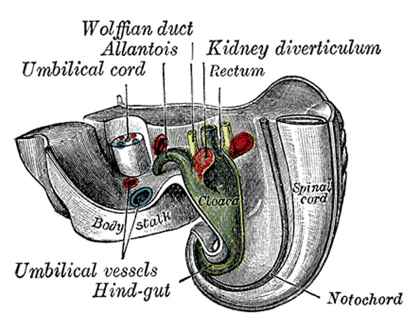
Tail end of Human Embryo 25 to 29 Days Old
(From model by Keibel.)
| Historic Disclaimer - information about historic embryology pages |
|---|
| Pages where the terms "Historic" (textbooks, papers, people, recommendations) appear on this site, and sections within pages where this disclaimer appears, indicate that the content and scientific understanding are specific to the time of publication. This means that while some scientific descriptions are still accurate, the terminology and interpretation of the developmental mechanisms reflect the understanding at the time of original publication and those of the preceding periods, these terms, interpretations and recommendations may not reflect our current scientific understanding. (More? Embryology History | Historic Embryology Papers) |
Metanephros and the Permanent Kidney
- rudiments of the permanent kidneys make their appearance about the end of the first or the beginning of the second month
- each kidney has a two-fold origin
- part arising from the metanephros
- part as a diverticulum from the hind-end of the Wolffian duct, close to where the latter opens into the cloaca
- metanephros arises in the intermediate cell mass caudal to the mesonephros, which it resembles in structure
- diverticulum from the Wolffian duct grows dorsalward and forward along the posterior abdominal wall
- its blind extremity expands and subsequently divides into several buds
- form the rudiments of the pelvis and calyces of the kidney
- by continued growth and subdivision it gives rise to the collecting tubules of the kidney
- proximal portion of the diverticulum becomes the ureter
- secretory tubules are developed from the metanephros, which is moulded over the growing end of the diverticulum from the Wolffian duct
(text modified from Gray's Anatomy)
- Gray's Images: Development | Lymphatic | Neural | Vision | Hearing | Somatosensory | Integumentary | Respiratory | Gastrointestinal | Urogenital | Endocrine | Surface Anatomy | iBook | Historic Disclaimer
| Historic Disclaimer - information about historic embryology pages |
|---|
| Pages where the terms "Historic" (textbooks, papers, people, recommendations) appear on this site, and sections within pages where this disclaimer appears, indicate that the content and scientific understanding are specific to the time of publication. This means that while some scientific descriptions are still accurate, the terminology and interpretation of the developmental mechanisms reflect the understanding at the time of original publication and those of the preceding periods, these terms, interpretations and recommendations may not reflect our current scientific understanding. (More? Embryology History | Historic Embryology Papers) |
| iBook - Gray's Embryology | |
|---|---|

|
|
Reference
Gray H. Anatomy of the human body. (1918) Philadelphia: Lea & Febiger.
Cite this page: Hill, M.A. (2024, April 27) Embryology Gray1115.jpg. Retrieved from https://embryology.med.unsw.edu.au/embryology/index.php/File:Gray1115.jpg
- © Dr Mark Hill 2024, UNSW Embryology ISBN: 978 0 7334 2609 4 - UNSW CRICOS Provider Code No. 00098G
File history
Click on a date/time to view the file as it appeared at that time.
| Date/Time | Thumbnail | Dimensions | User | Comment | |
|---|---|---|---|---|---|
| current | 08:39, 28 May 2011 |  | 600 × 474 (73 KB) | S8600021 (talk | contribs) | ==Tail end of Human Embryo 25 to 29 Days Old== (From model by Keibel.) {{Historic Disclaimer}} ===Metanephros and the Permanent Kidney=== * rudiments of the permanent kidneys make their appearance about the end of the first or the beginning of the se |
You cannot overwrite this file.
File usage
The following 5 pages use this file:

