File:Gray1114.jpg
From Embryology
Gray1114.jpg (450 × 471 pixels, file size: 47 KB, MIME type: image/jpeg)
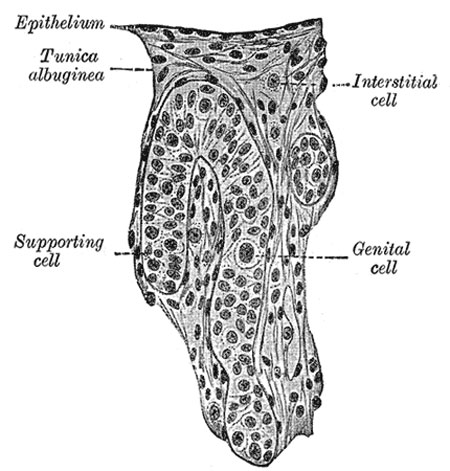
Human Embryo (3.5 cm long) Testis Section of a Genital Cord
(Felix and Bühler.)
| Historic Disclaimer - information about historic embryology pages |
|---|
| Pages where the terms "Historic" (textbooks, papers, people, recommendations) appear on this site, and sections within pages where this disclaimer appears, indicate that the content and scientific understanding are specific to the time of publication. This means that while some scientific descriptions are still accurate, the terminology and interpretation of the developmental mechanisms reflect the understanding at the time of original publication and those of the preceding periods, these terms, interpretations and recommendations may not reflect our current scientific understanding. (More? Embryology History | Historic Embryology Papers) |
The Testis
- testis is developed in much the same way as the ovary
- earliest stages it consists of a central mass of epithelium covered by a surface epithelium
- in the central mass a series of cords appear
- periphery of the mass is converted into the tunica albuginea
- excluding the surface epithelium from any part in the formation of the tissue of the testis
- cords of the central mass run together toward the future hilus
- form a network which ultimately becomes the rete testis
- cords develop the seminiferous tubules
- between them connective-tissue septa extend
- seminiferous tubules become connected with outgrowths from the Wolffian body
- form the efferent ducts of the testis
(text modified from Gray's Anatomy)
- Gray's Images: Development | Lymphatic | Neural | Vision | Hearing | Somatosensory | Integumentary | Respiratory | Gastrointestinal | Urogenital | Endocrine | Surface Anatomy | iBook | Historic Disclaimer
| Historic Disclaimer - information about historic embryology pages |
|---|
| Pages where the terms "Historic" (textbooks, papers, people, recommendations) appear on this site, and sections within pages where this disclaimer appears, indicate that the content and scientific understanding are specific to the time of publication. This means that while some scientific descriptions are still accurate, the terminology and interpretation of the developmental mechanisms reflect the understanding at the time of original publication and those of the preceding periods, these terms, interpretations and recommendations may not reflect our current scientific understanding. (More? Embryology History | Historic Embryology Papers) |
| iBook - Gray's Embryology | |
|---|---|

|
|
Reference
Gray H. Anatomy of the human body. (1918) Philadelphia: Lea & Febiger.
Cite this page: Hill, M.A. (2024, April 27) Embryology Gray1114.jpg. Retrieved from https://embryology.med.unsw.edu.au/embryology/index.php/File:Gray1114.jpg
- © Dr Mark Hill 2024, UNSW Embryology ISBN: 978 0 7334 2609 4 - UNSW CRICOS Provider Code No. 00098G
File history
Click on a date/time to view the file as it appeared at that time.
| Date/Time | Thumbnail | Dimensions | User | Comment | |
|---|---|---|---|---|---|
| current | 08:44, 28 May 2011 |  | 450 × 471 (47 KB) | S8600021 (talk | contribs) | ==Human Embryo (3.5 cm long) Testis Section of a Genital Cord== (Felix and Bühler.) {{Historic Disclaimer}} (text modified from Gray's Anatomy) {{Gray Anatomy}} Category:Human Category:Genital Category:Male Category:Testis |
You cannot overwrite this file.
File usage
The following 5 pages use this file:

