File:Gray0989.jpg
Original file (700 × 685 pixels, file size: 107 KB, MIME type: image/jpeg)
Week 8 Gastrointestinal Tract
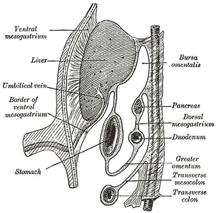
Schematic figure of the bursa omentalis, etc. Human embryo of eight weeks. (Kollmann.)
The lesser omentum is formed, as indicated above, by a thinning of the mesoderm or ventral mesogastrium, which attaches the stomach and duodenum to the anterior abdominal wall. By the subsequent growth of the liver this leaf of mesoderm is divided into two parts, viz., the lesser omentum between the stomach and liver, and the falciform and coronary ligaments between the liver and the abdominal wall and diaphragm (Fig. 989).
- Links: Image 987a | Image 987b | Image - Early Week 4 | Image - Late Week 4 | Gastrointestinal Tract Development | Endoderm
- Gray's Images: Development | Lymphatic | Neural | Vision | Hearing | Somatosensory | Integumentary | Respiratory | Gastrointestinal | Urogenital | Endocrine | Surface Anatomy | iBook | Historic Disclaimer
| Historic Disclaimer - information about historic embryology pages |
|---|
 |
| iBook - Gray's Embryology | |
|---|---|

|
|
Reference
Gray H. Anatomy of the human body. (1918) Philadelphia: Lea & Febiger.
Cite this page: Hill, M.A. (2024, April 27) Embryology Gray0989.jpg. Retrieved from https://embryology.med.unsw.edu.au/embryology/index.php/File:Gray0989.jpg
- © Dr Mark Hill 2024, UNSW Embryology ISBN: 978 0 7334 2609 4 - UNSW CRICOS Provider Code No. 00098G
File history
Click on a date/time to view the file as it appeared at that time.
| Date/Time | Thumbnail | Dimensions | User | Comment | |
|---|---|---|---|---|---|
| current | 15:19, 28 April 2011 | | 700 × 685 (107 KB) | S8600021 (talk | contribs) | ==Week 8 Gastrointestinal Tract== Schematic figure of the bursa omentalis, etc. Human embryo of eight weeks. (Kollmann.) :'''Links:''' Image 987a | Image 987b | [[:File:Gray0982a.jpg|Image - Early Week 4 |
You cannot overwrite this file.
File usage
The following page uses this file: