File:Gray0985.jpg
Gray0985.jpg (682 × 600 pixels, file size: 93 KB, MIME type: image/jpeg)
Gastrointestinal Tract Blood Supply
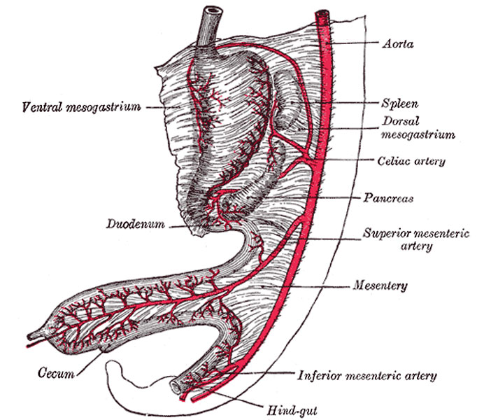
Abdominal part of digestive tube and its attachment to the primitive or common mesentery. Human embryo of six weeks. (After Toldt.)
With the lengthening of the tube, the mesoderm, which attaches it to the future vertebral column and carries the bloodvessels for the supply of the gut, is thinned and drawn out to form the posterior common mesentery. The portion of this mesentery attached to the greater curvature of the stomach is named the dorsal mesogastrium, and the part which suspends the colon is termed the mesocolon (Fig. 985). About the sixth week a diverticulum of the gut appears just behind the opening of the vitelline duct, and indicates the future cecum and vermiform process. The part of the loop on the distal side of the cecal diverticulum increases in diameter and forms the future ascending and transverse portions of the large intestine. Until the fifth month the cecal diverticulum has a uniform caliber, but from this time onward its distal part remains rudimentary and forms the vermiform process, while its proximal part expands to form the cecum.
Changes also take place in the shape and position of the stomach. Its dorsal part or greater curvature, to which the dorsal mesogastrium is attached, grows much more rapidly than its ventral part or lesser curvature to which the ventral mesogastrium is fixed. Further, the greater curvature is carried downward and to the left, so that the right surface of the stomach is now directed backward and the left surface forward (Fig. 986), a change in position which explains why the left vagus nerve is found on the front, and the right vagus on the back of the stomach. The dorsal mesogastrium being attached to the greater curvature must necessarily follow its movements, and hence it becomes greatly elongated and drawn lateralward and ventralward from the vertebral column, and, as in the case of the stomach, the right surfaces of both the dorsal and ventral mesogastria are now directed backward, and the left forward. In this way a pouch, the bursa omentalis, is formed behind the stomach, and this increases in size as the digestive tube undergoes further development; the entrance to the pouch constitutes the future foramen epiploicum or foramen of Winslow.
The duodenum is developed from that part of the tube which immediately succeeds the stomach; it undergoes little elongation, being more or less fixed in position by the liver and pancreas, which arise as diverticula from it. The duodenum is at first suspended by a mesentery, and projects forward in the form of a loop. The loop and its mesentery are subsequently displaced by the transverse colon, so that the right surface of the duodenal mesentery is directed backward, and, adhering to the parietal peritoneum, is lost. The remainder of the digestive tube becomes greatly elongated, and as a consequence the tube is coiled on itself, and this elongation demands a corresponding increase in the width of the intestinal attachment of the mesentery, which becomes folded.
- Gray's Images: Development | Lymphatic | Neural | Vision | Hearing | Somatosensory | Integumentary | Respiratory | Gastrointestinal | Urogenital | Endocrine | Surface Anatomy | iBook | Historic Disclaimer
| Historic Disclaimer - information about historic embryology pages |
|---|
| Pages where the terms "Historic" (textbooks, papers, people, recommendations) appear on this site, and sections within pages where this disclaimer appears, indicate that the content and scientific understanding are specific to the time of publication. This means that while some scientific descriptions are still accurate, the terminology and interpretation of the developmental mechanisms reflect the understanding at the time of original publication and those of the preceding periods, these terms, interpretations and recommendations may not reflect our current scientific understanding. (More? Embryology History | Historic Embryology Papers) |
| iBook - Gray's Embryology | |
|---|---|

|
|
Reference
Gray H. Anatomy of the human body. (1918) Philadelphia: Lea & Febiger.
Cite this page: Hill, M.A. (2024, April 27) Embryology Gray0985.jpg. Retrieved from https://embryology.med.unsw.edu.au/embryology/index.php/File:Gray0985.jpg
- © Dr Mark Hill 2024, UNSW Embryology ISBN: 978 0 7334 2609 4 - UNSW CRICOS Provider Code No. 00098G
File history
Click on a date/time to view the file as it appeared at that time.
| Date/Time | Thumbnail | Dimensions | User | Comment | |
|---|---|---|---|---|---|
| current | 23:40, 21 August 2012 |  | 682 × 600 (93 KB) | Z8600021 (talk | contribs) |
You cannot overwrite this file.
File usage
The following page uses this file:

