File:Gray0983.jpg
Gray0983.jpg (800 × 517 pixels, file size: 38 KB, MIME type: image/jpeg)
Week 4-5 - Early Human Digestive Tube
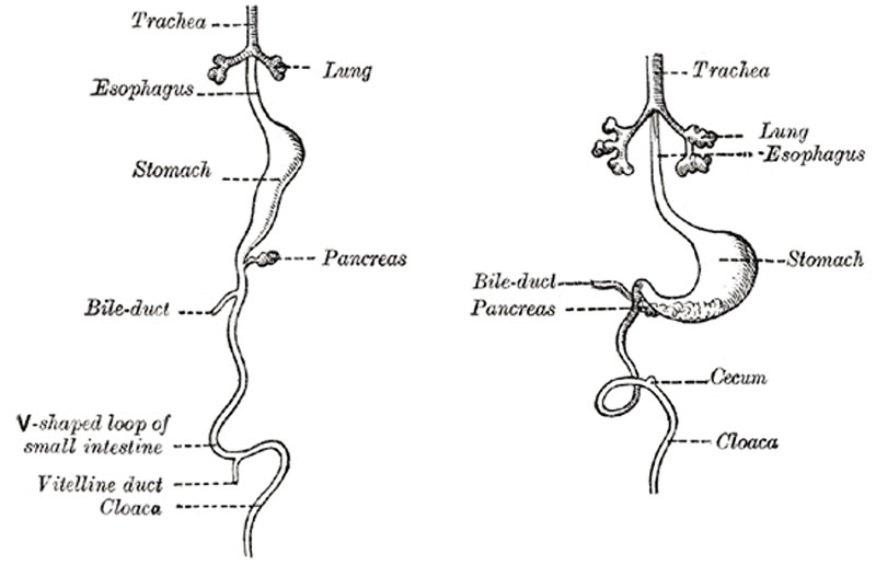
Front view of two successive stages in the development of the digestive tube. (His.) (See enlarged image)
About the fourth week a fusiform dilatation, the future stomach, makes its appearance, and beyond this the gut opens freely into the yolk-sac (Fig. 982, A and B). The opening is at first wide, but is gradually narrowed into a tubular stalk, the yolk-stalk or vitelline duct. Between the stomach and the mouth of the yolk-sac the liver diverticulum appears. From the stomach to the rectum the alimentary canal is attached to the notochord by a band of mesoderm, from which the common mesentery of the gut is subsequently developed. The stomach has an additional attachment, viz., to the ventral abdominal wall as far as the umbilicus by the septum transversum. The cephalic portion of the septum takes part in the formation of the diaphragm, while the caudal portion into which the liver grows forms the ventral mesogastrium.
The stomach undergoes a further dilatation, and its two curvatures can be recognized (Figs. 983, B, and 984), the greater directed toward the vertebral column and the lesser toward the anterior wall of the abdomen, while its two surfaces look to the right and left respectively. Behind the stomach the gut undergoes great elongation, and forms a V-shaped loop which projects downward and forward; from the bend or angle of the loop the vitelline duct passes to the umbilicus (Fig. 984).
- Gray's Images: Development | Lymphatic | Neural | Vision | Hearing | Somatosensory | Integumentary | Respiratory | Gastrointestinal | Urogenital | Endocrine | Surface Anatomy | iBook | Historic Disclaimer
| Historic Disclaimer - information about historic embryology pages |
|---|
| Pages where the terms "Historic" (textbooks, papers, people, recommendations) appear on this site, and sections within pages where this disclaimer appears, indicate that the content and scientific understanding are specific to the time of publication. This means that while some scientific descriptions are still accurate, the terminology and interpretation of the developmental mechanisms reflect the understanding at the time of original publication and those of the preceding periods, these terms, interpretations and recommendations may not reflect our current scientific understanding. (More? Embryology History | Historic Embryology Papers) |
| iBook - Gray's Embryology | |
|---|---|

|
|
Reference
Gray H. Anatomy of the human body. (1918) Philadelphia: Lea & Febiger.
Cite this page: Hill, M.A. (2024, April 27) Embryology Gray0983.jpg. Retrieved from https://embryology.med.unsw.edu.au/embryology/index.php/File:Gray0983.jpg
- © Dr Mark Hill 2024, UNSW Embryology ISBN: 978 0 7334 2609 4 - UNSW CRICOS Provider Code No. 00098G
File history
Click on a date/time to view the file as it appeared at that time.
| Date/Time | Thumbnail | Dimensions | User | Comment | |
|---|---|---|---|---|---|
| current | 14:38, 28 April 2011 |  | 800 × 517 (38 KB) | S8600021 (talk | contribs) | ==Early Human Digestive Tube== Front view of two successive stages in the development of the digestive tube. (His.) (See enlarged image) About the fourth week a fusiform dilatation, the future stomach, makes its appearance, and beyond this the gut opens |
You cannot overwrite this file.
File usage
The following page uses this file:

