File:Gray0978.jpg
Gray0978.jpg (483 × 600 pixels, file size: 67 KB, MIME type: image/jpeg)
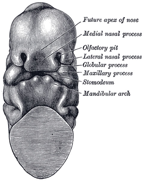
Head end of human embryo of about thirty to thirty-one days
(From model by Peters.)
The Mouth
The mouth is developed partly from the stomodeum, and partly from the floor of the anterior portion of the fore-gut. By the growth of the head end of the embryo, and the formation of the cephalic flexure, the pericardial area and the buccopharyngeal membrane come to lie on the ventral surface of the embryo. With the further expansion of the brain, and the forward bulging of the pericardium, the buccopharyngeal membrane is depressed between these two prominences. This depression constitutes the stomodeum (Fig. 977). It is lined by ectoderm, and is separated from the anterior end of the fore-gut by the buccopharyngeal membrane. This membrane is devoid of mesoderm, being formed by the apposition of the stomodeal ectoderm with the fore-gut entoderm; at the end of the third week it disappears, and thus a communication is established between the mouth and the future pharynx. No trace of the membrane is found in the adult; and the communication just mentioned must not be confused with the permanent isthmus faucium. The lips, teeth, and gums are formed from the walls of the stomodeum, but the tongue is developed in the floor of the pharynx.
The visceral arches extend in a ventral direction between the stomodeum and the pericardium; and with the completion of the mandibular arch and the formation of the maxillary processes, the mouth assumes the appearance of a pentagonal orifice. The orifice is bounded in front by the fronto-nasal process, behind by the mandibular arch, and laterally by the maxillary processes (Fig. 978). With the inward growth and fusion of the palatine processes (Figs. 50, 51), the stomodeum is divided into an upper nasal, and a lower buccal part. Along the free margins of the processes bounding the mouth cavity a shallow groove appears; this is termed the primary labial groove, and from the bottom of it a downgrowth of ectoderm takes place into the underlying mesoderm. The central cells of the ectodermal downgrowth degenerate and a secondary labial groove is formed; by the deepening of this, the lips and cheeks are separated from the alveolar processes of the maxillæ and mandible.
- Gray's Images: Development | Lymphatic | Neural | Vision | Hearing | Somatosensory | Integumentary | Respiratory | Gastrointestinal | Urogenital | Endocrine | Surface Anatomy | iBook | Historic Disclaimer
| Historic Disclaimer - information about historic embryology pages |
|---|
| Pages where the terms "Historic" (textbooks, papers, people, recommendations) appear on this site, and sections within pages where this disclaimer appears, indicate that the content and scientific understanding are specific to the time of publication. This means that while some scientific descriptions are still accurate, the terminology and interpretation of the developmental mechanisms reflect the understanding at the time of original publication and those of the preceding periods, these terms, interpretations and recommendations may not reflect our current scientific understanding. (More? Embryology History | Historic Embryology Papers) |
| iBook - Gray's Embryology | |
|---|---|

|
|
Reference
Gray H. Anatomy of the human body. (1918) Philadelphia: Lea & Febiger.
Cite this page: Hill, M.A. (2024, April 27) Embryology Gray0978.jpg. Retrieved from https://embryology.med.unsw.edu.au/embryology/index.php/File:Gray0978.jpg
- © Dr Mark Hill 2024, UNSW Embryology ISBN: 978 0 7334 2609 4 - UNSW CRICOS Provider Code No. 00098G
File history
Click on a date/time to view the file as it appeared at that time.
| Date/Time | Thumbnail | Dimensions | User | Comment | |
|---|---|---|---|---|---|
| current | 00:08, 22 August 2012 |  | 483 × 600 (67 KB) | Z8600021 (talk | contribs) | {{Gray Anatomy}} Category:Gastrointestinal Tract Category:Human Category:Pharyngeal Arch Category:Tongue |
You cannot overwrite this file.
File usage
The following page uses this file:

