File:Gray0945.jpg
Gray0945.jpg (400 × 585 pixels, file size: 76 KB, MIME type: image/jpeg)
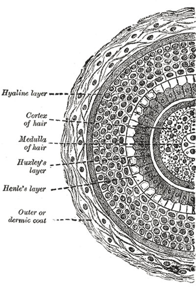
Transverse section of hair follicle
The outer or dermic coat is formed mainly of fibrous tissue; it is continuous with the corium, is highly vascular, and supplied by numerous minute nervous filaments. It consists of three layers (Fig. 945). The most internal is a hyaline basement membrane, which is well-marked in the larger hair follicles, but is not very distinct in the follicles of minute hairs; it is limited to the deeper part of the follicle. Outside this is a compact layer of fibers and spindle-shaped cells arranged circularly around the follicle; this layer extends from the bottom of the follicle as high as the entrance of the ducts of the sebaceous glands. Externally is a thick layer of connective tissue, arranged in longitudinal bundles, forming a more open texture and corresponding to the reticular part of the corium; in this are contained the bloodvessels and nerves.
- Gray's Images: Development | Lymphatic | Neural | Vision | Hearing | Somatosensory | Integumentary | Respiratory | Gastrointestinal | Urogenital | Endocrine | Surface Anatomy | iBook | Historic Disclaimer
| Historic Disclaimer - information about historic embryology pages |
|---|
| Pages where the terms "Historic" (textbooks, papers, people, recommendations) appear on this site, and sections within pages where this disclaimer appears, indicate that the content and scientific understanding are specific to the time of publication. This means that while some scientific descriptions are still accurate, the terminology and interpretation of the developmental mechanisms reflect the understanding at the time of original publication and those of the preceding periods, these terms, interpretations and recommendations may not reflect our current scientific understanding. (More? Embryology History | Historic Embryology Papers) |
| iBook - Gray's Embryology | |
|---|---|

|
|
Reference
Gray H. Anatomy of the human body. (1918) Philadelphia: Lea & Febiger.
Cite this page: Hill, M.A. (2024, April 27) Embryology Gray0945.jpg. Retrieved from https://embryology.med.unsw.edu.au/embryology/index.php/File:Gray0945.jpg
- © Dr Mark Hill 2024, UNSW Embryology ISBN: 978 0 7334 2609 4 - UNSW CRICOS Provider Code No. 00098G
File history
Click on a date/time to view the file as it appeared at that time.
| Date/Time | Thumbnail | Dimensions | User | Comment | |
|---|---|---|---|---|---|
| current | 23:51, 19 August 2012 |  | 400 × 585 (76 KB) | Z8600021 (talk | contribs) | ==Transverse section of hair follicle== The outer or dermic coat is formed mainly of fibrous tissue; it is continuous with the corium, is highly vascular, and supplied by numerous minute nervous filaments. It consists of three layers (Fig. 945). The mos |
You cannot overwrite this file.
File usage
The following page uses this file:

