File:Gray0944.jpg

Original file (800 × 681 pixels, file size: 178 KB, MIME type: image/jpeg)
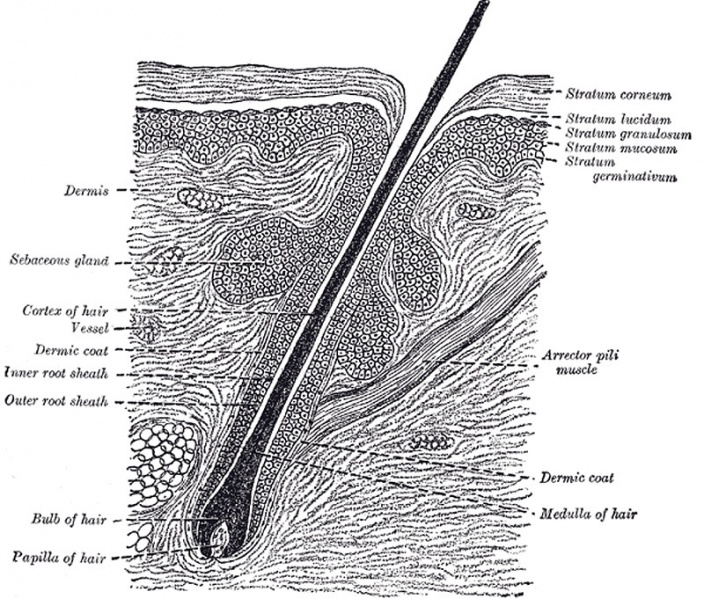
Section of skin
Section of skin, showing the epidermis and dermis; a hair in its follicle; the Arrector pili muscle; sebaceous glands.
Hairs (pili) are found on nearly every part of the surface of the body, but are absent from the palms of the hands, the soles of the feet, the dorsal surfaces of the terminal phalanges, the glans penis, the inner surface of the prepuce, and the inner surfaces of the labia. They vary much in length, thickness, and color in different parts of the body and in different races of mankind. In some parts, as in the skin of the eyelids, they are so short as not to project beyond the follicles containing them; in others, as upon the scalp, they are of considerable length; again, in other parts, as the eyelashes, the hairs of the pubic region, and the whiskers and beard, they are remarkable for their thickness. Straight hairs are stronger than curly hairs, and present on transverse section a cylindrical or oval outline; curly hairs, on the other hand, are flattened. A hair consists of a root, the part implanted in the skin; and a shaft or scapus, the portion projecting from the surface.
The root of the hair (radix pili) ends in an enlargement, the hair bulb, which is whiter in color and softer in texture than the shaft, and is lodged in a follicular involution of the epidermis called the hair follicle (Fig. 944). When the hair is of considerable length the follicle extends into the subcutaneous cellular tissue. The hair follicle commences on the surface of the skin with a funnel-shaped opening, and passes inward in an oblique or curved direction—the latter in curly hairs—to become dilated at its deep extremity, where it corresponds with the hair bulb. Opening into the follicle, near its free extremity, are the ducts of one or more sebaceous glands. At the bottom of each hair follicle is a small conical, vascular eminence or papilla, similar in every respect to those found upon the surface of the skin; it is continuous with the dermic layer of the follicle, and is supplied with nerve fibrils. The hair follicle consists of two coats—an outer or dermic, and an inner or epidermic.
- Gray's Images: Development | Lymphatic | Neural | Vision | Hearing | Somatosensory | Integumentary | Respiratory | Gastrointestinal | Urogenital | Endocrine | Surface Anatomy | iBook | Historic Disclaimer
| Historic Disclaimer - information about historic embryology pages |
|---|
| Pages where the terms "Historic" (textbooks, papers, people, recommendations) appear on this site, and sections within pages where this disclaimer appears, indicate that the content and scientific understanding are specific to the time of publication. This means that while some scientific descriptions are still accurate, the terminology and interpretation of the developmental mechanisms reflect the understanding at the time of original publication and those of the preceding periods, these terms, interpretations and recommendations may not reflect our current scientific understanding. (More? Embryology History | Historic Embryology Papers) |
| iBook - Gray's Embryology | |
|---|---|

|
|
Reference
Gray H. Anatomy of the human body. (1918) Philadelphia: Lea & Febiger.
Cite this page: Hill, M.A. (2024, April 27) Embryology Gray0944.jpg. Retrieved from https://embryology.med.unsw.edu.au/embryology/index.php/File:Gray0944.jpg
- © Dr Mark Hill 2024, UNSW Embryology ISBN: 978 0 7334 2609 4 - UNSW CRICOS Provider Code No. 00098G
File history
Click on a date/time to view the file as it appeared at that time.
| Date/Time | Thumbnail | Dimensions | User | Comment | |
|---|---|---|---|---|---|
| current | 23:49, 19 August 2012 |  | 800 × 681 (178 KB) | Z8600021 (talk | contribs) | Section of skin, showing the epidermis and dermis; a hair in its follicle; the Arrector pili muscle; sebaceous glands. Hairs (pili) are found on nearly every part of the surface of the body, but are absent from the palms of the hands, the soles of the f |
You cannot overwrite this file.
File usage
The following page uses this file:
