File:Gray0943.jpg
From Embryology
Gray0943.jpg (800 × 277 pixels, file size: 71 KB, MIME type: image/jpeg)
The Nails
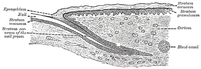
The Nails (ungues) are flattened, elastic structures of a horny texture, placed upon the dorsal surfaces of the terminal phalanges of the fingers and toes.
- Each nail is convex on its outer surface, concave within, and is implanted by a portion, called the root, into a groove in the skin; the exposed portion is called the body, and the distal extremity the free edge.
- The nail is firmly adherent to the corium, being accurately moulded upon its surface; the part beneath the body and root of the nail is called the nail matrix, because from it the nail is produced.
- Under the greater part of the body of the nail, the matrix is thick, and raised into a series of longitudinal ridges which are very vascular, and the color is seen through the transparent tissue.
- Near the root of the nail, the papillæ are smaller, less vascular, and have no regular arrangement, and here the tissue of the nail is not firmly adherent to the connective-tissue stratum but only in contact with it; hence this portion is of a whiter color, and is called the lunula on account of its shape.
- The cuticle as it passes forward on the dorsal surface of the finger or toe is attached to the surface of the nail a little in advance of its root; at the extremity of the finger it is connected with the under surface of the nail a little behind its free edge.
- The cuticle and the horny substance of the nail (both epidermic structures) are thus directly continuous with each other.
- The superficial, horny part of the nail consists of a greatly thickened stratum lucidum, the stratum corneum forming merely the thin cuticular fold (eponychium) which overlaps the lunula; the deeper part consists of the stratum mucosum.
- The cells in contact with the papillæ of the matrix are columnar in form and arranged perpendicularly to the surface; those which succeed them are of a rounded or polygonal form, the more superficial ones becoming broad, thin, and flattened, and so closely packed as to make the limits of the cells very indistinct.
- The nails grow in length by the proliferation of the cells of the stratum mucosum at the root of the nail, and in thickness from that part of the stratum mucosum which underlies the lunula.
- Gray's Images: Development | Lymphatic | Neural | Vision | Hearing | Somatosensory | Integumentary | Respiratory | Gastrointestinal | Urogenital | Endocrine | Surface Anatomy | iBook | Historic Disclaimer
| Historic Disclaimer - information about historic embryology pages |
|---|
| Pages where the terms "Historic" (textbooks, papers, people, recommendations) appear on this site, and sections within pages where this disclaimer appears, indicate that the content and scientific understanding are specific to the time of publication. This means that while some scientific descriptions are still accurate, the terminology and interpretation of the developmental mechanisms reflect the understanding at the time of original publication and those of the preceding periods, these terms, interpretations and recommendations may not reflect our current scientific understanding. (More? Embryology History | Historic Embryology Papers) |
| iBook - Gray's Embryology | |
|---|---|

|
|
Reference
Gray H. Anatomy of the human body. (1918) Philadelphia: Lea & Febiger.
Cite this page: Hill, M.A. (2024, April 27) Embryology Gray0943.jpg. Retrieved from https://embryology.med.unsw.edu.au/embryology/index.php/File:Gray0943.jpg
- © Dr Mark Hill 2024, UNSW Embryology ISBN: 978 0 7334 2609 4 - UNSW CRICOS Provider Code No. 00098G
File history
Click on a date/time to view the file as it appeared at that time.
| Date/Time | Thumbnail | Dimensions | User | Comment | |
|---|---|---|---|---|---|
| current | 01:40, 21 September 2010 | 800 × 277 (71 KB) | S8600021 (talk | contribs) | ==The Nails== The Nails (ungues) (Fig. 943) are flattened, elastic structures of a horny texture, placed upon the dorsal surfaces of the terminal phalanges of the fingers and toes. Each nail is convex on its outer surface, concave within, and is implante |
You cannot overwrite this file.
File usage
The following 3 pages use this file:

