File:Gray0941.jpg
Gray0941.jpg (700 × 524 pixels, file size: 106 KB, MIME type: image/jpeg)
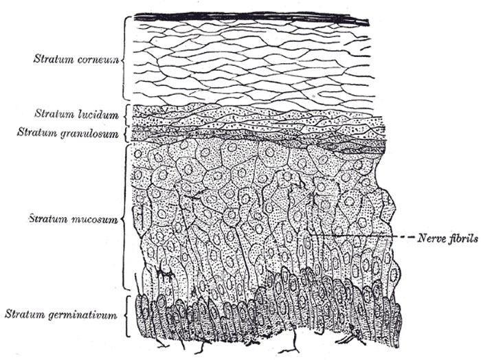
Section of Epidermis
(Ranvier.)
The epidermis, cuticle, or scarf skin is non-vascular, and consists of stratified epithelium (Fig. 941), and is accurately moulded on the papillary layer of the corium. It varies in thickness in different parts. In some situations, as in the palms of the hands and soles of the feet, it is thick, hard, and horny in texture. This may be in a measure due to the fact that these parts are exposed to intermittent pressure, but that this is not the only cause is proved by the fact that the condition exists to a very considerable extent at birth. The more superficial layers of cells, called the horny layer (stratum corneum), may be separated by maceration from a deeper stratum, which is called the stratum mucosum, and which consists of several layers of differently shaped cells. The free surface of the epidermis is marked by a net-work of linear furrows of variable size, dividing the surface into a number of polygonal or lozenge-shaped areas. Some of these furrows are large, as opposite the flexures of the joints, and correspond to the folds in the corium produced by movements. In other situations, as upon the back of the hand, they are exceedingly fine, and intersect one another at various angles. Upon the palmar surfaces of the hands and fingers, and upon the soles of the feet, the epidermal ridges are very distinct, and are disposed in curves; they depend upon the large size and peculiar arrangements of the papillæ upon which the epidermis is placed. The function of these ridges is primarily to increase resistance between contact surfaces for the purpose of preventing slipping whether in walking or prehension. The direction of the ridges is at right angles with the force that tends to produce slipping or to the resultant of such forces when these forces vary in direction. 153 In each individual the lines on the tips of the fingers and thumbs form distinct patterns unlike those of any other person. A method of determining the identity of a criminal is based on this fact, impressions (“finger-prints”) of these lines being made on paper covered with soot, or on white paper after first covering the fingers with ink. The deep surface of the epidermis is accurately moulded upon the papillary layer of the corium, the papillæ being covered by a basement membrane; so that when the epidermis is removed by maceration, it presents on its under surface a number of pits or depressions corresponding to the papillæ, and ridges corresponding to the intervals between them. Fine tubular prolongations are continued from this layer into the ducts of the sudoriferous and sebaceous glands.
(Text modified from Gray's 1918 Anatomy)
- Gray's Images: Development | Lymphatic | Neural | Vision | Hearing | Somatosensory | Integumentary | Respiratory | Gastrointestinal | Urogenital | Endocrine | Surface Anatomy | iBook | Historic Disclaimer
| Historic Disclaimer - information about historic embryology pages |
|---|
| Pages where the terms "Historic" (textbooks, papers, people, recommendations) appear on this site, and sections within pages where this disclaimer appears, indicate that the content and scientific understanding are specific to the time of publication. This means that while some scientific descriptions are still accurate, the terminology and interpretation of the developmental mechanisms reflect the understanding at the time of original publication and those of the preceding periods, these terms, interpretations and recommendations may not reflect our current scientific understanding. (More? Embryology History | Historic Embryology Papers) |
| iBook - Gray's Embryology | |
|---|---|

|
|
Reference
Gray H. Anatomy of the human body. (1918) Philadelphia: Lea & Febiger.
Cite this page: Hill, M.A. (2024, April 27) Embryology Gray0941.jpg. Retrieved from https://embryology.med.unsw.edu.au/embryology/index.php/File:Gray0941.jpg
- © Dr Mark Hill 2024, UNSW Embryology ISBN: 978 0 7334 2609 4 - UNSW CRICOS Provider Code No. 00098G
File history
Click on a date/time to view the file as it appeared at that time.
| Date/Time | Thumbnail | Dimensions | User | Comment | |
|---|---|---|---|---|---|
| current | 23:38, 19 August 2012 |  | 700 × 524 (106 KB) | Z8600021 (talk | contribs) | ==Section of Epidermis== (Ranvier.) The epidermis, cuticle, or scarf skin is non-vascular, and consists of stratified epithelium (Fig. 941), and is accurately moulded on the papillary layer of the corium. It varies in thickness in different parts. In so |
You cannot overwrite this file.
File usage
The following page uses this file:

