File:Gray0939.jpg
Gray0939.jpg (681 × 371 pixels, file size: 90 KB, MIME type: image/jpeg)
Muscle Spindle
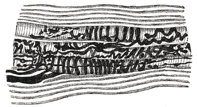
Middle third of a terminal plaque in the muscle spindle of an adult cat. (After Ruffini.)
The neuromuscular spindles are present in the majority of voluntary muscles, and consist of small bundles of peculiar muscular fibers (intrafusal fibers), embryonic in type, invested by capsules, within which nerve fibers, experimentally shown to be sensory in origin, terminate. These neuromuscular spindles vary in length from 0.8 mm. to 5 mm., and have a distinctly fusiform appearance. The large medullated nerve fibers passing to the end-organ are from one to three or four in number; entering the fibrous capsule, they divide several times, and, losing their medullary sheaths, ultimately end in naked axis-cylinders encircling the intrafusal fibers by flattened expansions, or irregular ovoid or rounded disks (Fig. 939). Neuromuscular spindles have not yet been demonstrated in the tongue muscles, and only a few exist in the ocular muscles.
(Text modified from Gray's 1918 Anatomy)
- Gray's Images: Development | Lymphatic | Neural | Vision | Hearing | Somatosensory | Integumentary | Respiratory | Gastrointestinal | Urogenital | Endocrine | Surface Anatomy | iBook | Historic Disclaimer
| Historic Disclaimer - information about historic embryology pages |
|---|
| Pages where the terms "Historic" (textbooks, papers, people, recommendations) appear on this site, and sections within pages where this disclaimer appears, indicate that the content and scientific understanding are specific to the time of publication. This means that while some scientific descriptions are still accurate, the terminology and interpretation of the developmental mechanisms reflect the understanding at the time of original publication and those of the preceding periods, these terms, interpretations and recommendations may not reflect our current scientific understanding. (More? Embryology History | Historic Embryology Papers) |
| iBook - Gray's Embryology | |
|---|---|

|
|
Reference
Gray H. Anatomy of the human body. (1918) Philadelphia: Lea & Febiger.
Cite this page: Hill, M.A. (2024, April 27) Embryology Gray0939.jpg. Retrieved from https://embryology.med.unsw.edu.au/embryology/index.php/File:Gray0939.jpg
- © Dr Mark Hill 2024, UNSW Embryology ISBN: 978 0 7334 2609 4 - UNSW CRICOS Provider Code No. 00098G
File history
Click on a date/time to view the file as it appeared at that time.
| Date/Time | Thumbnail | Dimensions | User | Comment | |
|---|---|---|---|---|---|
| current | 08:35, 19 August 2012 |  | 681 × 371 (90 KB) | Z8600021 (talk | contribs) | ==Muscle Spindle== Middle third of a terminal plaque in the muscle spindle of an adult cat. (After Ruffini.) The neuromuscular spindles are present in the majority of voluntary muscles, and consist of small bundles of peculiar muscular fibers (intrafus |
You cannot overwrite this file.
File usage
The following page uses this file:

