File:Gray0896.jpg

Original file (600 × 638 pixels, file size: 68 KB, MIME type: image/jpeg)
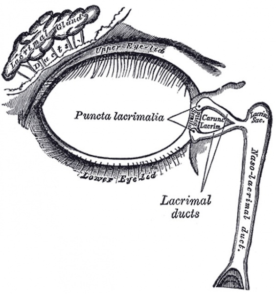
The lacrimal apparatus
Right side.
The Lacrimal Apparatus (apparatus lacrimalis) (Fig. 896) consists of (a) the lacrimal gland, which secretes the tears, and its excretory ducts, which convey the fluid to the surface of the eye; (b) the lacrimal ducts, the lacrimal sac, and the nasolacrimal duct, by which the fluid is conveyed into the cavity of the nose.
The Lacrimal Gland (glandula lacrimalis).—The lacrimal gland is lodged in the lacrimal fossa, on the medial side of the zygomatic process of the frontal bone. It is of an oval form, about the size and shape of an almond, and consists of two portions, described as the superior and inferior lacrimal glands. The superior lacrimal gland is connected to the periosteum of the orbit by a few fibrous bands, and rests upon the tendons of the Recti superioris and lateralis, which separate it from the bulb of the eye. The inferior lacrimal gland is separated from the superior by a fibrous septum, and projects into the back part of the upper eyelid, where its deep surface is related to the conjunctiva. The ducts of the glands, from six to twelve in number, run obliquely beneath the conjunctiva for a short distance, and open along the upper and lateral half of the superior conjunctival fornix.
(Text modified from Gray's 1918 Anatomy)
- Gray's Images: Development | Lymphatic | Neural | Vision | Hearing | Somatosensory | Integumentary | Respiratory | Gastrointestinal | Urogenital | Endocrine | Surface Anatomy | iBook | Historic Disclaimer
| Historic Disclaimer - information about historic embryology pages |
|---|
| Pages where the terms "Historic" (textbooks, papers, people, recommendations) appear on this site, and sections within pages where this disclaimer appears, indicate that the content and scientific understanding are specific to the time of publication. This means that while some scientific descriptions are still accurate, the terminology and interpretation of the developmental mechanisms reflect the understanding at the time of original publication and those of the preceding periods, these terms, interpretations and recommendations may not reflect our current scientific understanding. (More? Embryology History | Historic Embryology Papers) |
| iBook - Gray's Embryology | |
|---|---|

|
|
Reference
Gray H. Anatomy of the human body. (1918) Philadelphia: Lea & Febiger.
Cite this page: Hill, M.A. (2024, April 27) Embryology Gray0896.jpg. Retrieved from https://embryology.med.unsw.edu.au/embryology/index.php/File:Gray0896.jpg
- © Dr Mark Hill 2024, UNSW Embryology ISBN: 978 0 7334 2609 4 - UNSW CRICOS Provider Code No. 00098G
File history
Click on a date/time to view the file as it appeared at that time.
| Date/Time | Thumbnail | Dimensions | User | Comment | |
|---|---|---|---|---|---|
| current | 23:03, 19 August 2012 |  | 600 × 638 (68 KB) | Z8600021 (talk | contribs) | ==The lacrimal apparatus== Right side. The Lacrimal Apparatus (apparatus lacrimalis) (Fig. 896) consists of (a) the lacrimal gland, which secretes the tears, and its excretory ducts, which convey the fluid to the surface of the eye; (b) the lacrimal duc |
You cannot overwrite this file.
File usage
The following 2 pages use this file:
