File:Gray0891.jpg
Gray0891.jpg (600 × 544 pixels, file size: 104 KB, MIME type: image/jpeg)
Right Eye Fascia Bulbi
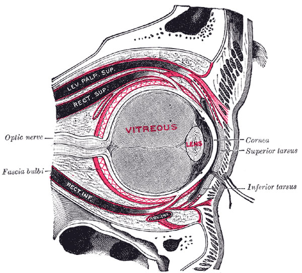
The right eye in sagittal section, showing the fascia bulbi (semidiagrammatic). (Testut.)
The Fascia Bulb (capsule of Ténon) (Fig. 891) is a thin membrane which envelops the bulb of the eye from the optic nerve to the ciliary region, separating it from the orbital fat and forming a socket in which it plays. Its inner surface is smooth, and is separated from the outer surface of the sclera by the periscleral lymph space. This lymph space is continuous with the subdural and subarachnoid cavities, and is traversed by delicate bands of connective tissue which extend between the fascia and the sclera. The fascia is perforated behind by the ciliary vessels and nerves, and fuses with the sheath of the optic nerve and with the sclera around the entrance of the optic nerve. In front it blends with the ocular conjunctiva, and with it is attached to the ciliary region of the eyeball. It is perforated by the tendons of the ocular muscles, and is reflected backward on each as a tubular sheath. The sheath of the Obliquus superior is carried as far as the fibrous pulley of that muscle; that on the Obliquus inferior reaches as far as the floor of the orbit, to which it gives off a slip. The sheaths on the Recti are gradually lost in the perimysium, but they give off important expansions. The expansion from the Rectus superior blends with the tendon of the Levator palpebræ; that of the Rectus inferior is attached to the inferior tarsus. The expansions from the sheaths of the Recti lateralis and medialis are strong, especially that from the latter muscle, and are attached to the lacrimal and zygomatic bones respectively. As they probably check the actions of these two Recti they have been named the medial and lateral check ligaments. Lockwood has described a thickening of the lower part of the facia bulbi, which he has named the suspensory ligament of the eye. It is slung like a hammock below the eyeball, being expanded in the center, and narrow at its extremities which are attached to the zygomatic and lacrimal bones respectively.
The Orbital Fascia forms the periosteum of the orbit. It is loosely connected to the bones and can be readily separated from them. Behind, it is united with the dura mater by processes which pass through the optic foramen and superior orbital fissure, and with the sheath of the optic nerve. In front, it is connected with the periosteum at the margin of the orbit, and sends off a process which assists in forming the orbital septum. From it two processes are given off; one to enclose the lacrimal gland, the other to hold the pulley of the Obliquus superior in position.
(Text modified from Gray's 1918 Anatomy)
- Gray's Images: Development | Lymphatic | Neural | Vision | Hearing | Somatosensory | Integumentary | Respiratory | Gastrointestinal | Urogenital | Endocrine | Surface Anatomy | iBook | Historic Disclaimer
| Historic Disclaimer - information about historic embryology pages |
|---|
| Pages where the terms "Historic" (textbooks, papers, people, recommendations) appear on this site, and sections within pages where this disclaimer appears, indicate that the content and scientific understanding are specific to the time of publication. This means that while some scientific descriptions are still accurate, the terminology and interpretation of the developmental mechanisms reflect the understanding at the time of original publication and those of the preceding periods, these terms, interpretations and recommendations may not reflect our current scientific understanding. (More? Embryology History | Historic Embryology Papers) |
| iBook - Gray's Embryology | |
|---|---|

|
|
Reference
Gray H. Anatomy of the human body. (1918) Philadelphia: Lea & Febiger.
Cite this page: Hill, M.A. (2024, April 27) Embryology Gray0891.jpg. Retrieved from https://embryology.med.unsw.edu.au/embryology/index.php/File:Gray0891.jpg
- © Dr Mark Hill 2024, UNSW Embryology ISBN: 978 0 7334 2609 4 - UNSW CRICOS Provider Code No. 00098G
File history
Click on a date/time to view the file as it appeared at that time.
| Date/Time | Thumbnail | Dimensions | User | Comment | |
|---|---|---|---|---|---|
| current | 22:48, 19 August 2012 |  | 600 × 544 (104 KB) | Z8600021 (talk | contribs) | ==Right Eye Fascia Bulbi== The right eye in sagittal section, showing the fascia bulbi (semidiagrammatic). (Testut.) The Fascia Bulb (capsule of Ténon) (Fig. 891) is a thin membrane which envelops the bulb of the eye from the optic nerve to the ciliar |
You cannot overwrite this file.
File usage
The following page uses this file:

