File:Gray0884.jpg
Gray0884.jpg (419 × 400 pixels, file size: 58 KB, MIME type: image/jpeg)
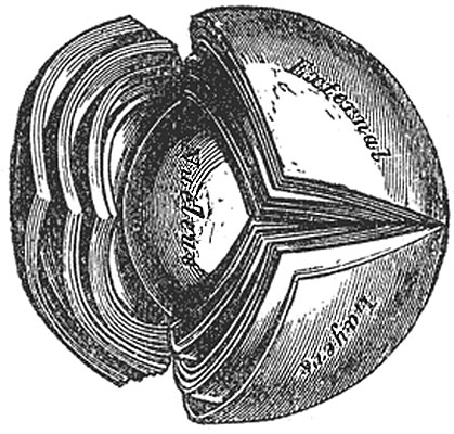
Lens Structure
The crystalline lens, hardened and divided. (Enlarged.)
The lens is made up of soft cortical substance and a firm, central part, the nucleus (Fig. 884). Faint lines (radii lentis) radiate from the poles to the equator. In the adult there may be six or more of these lines, but in the fetus they are only three in number and diverge from each other at angles of 120° (Fig. 885); on the anterior surface one line ascends vertically and the other two diverge downward; on the posterior surface one ray descends vertically and the other two diverge upward. They correspond with the free edges of an equal number of septa composed of an amorphous substance, which dip into the substance of the lens. When the lens has been hardened it is seen to consist of a series of concentrically arranged laminæ, each of which is interrupted at the septa referred to. Each lamina is built up of a number of hexagonal, ribbon-like lens fibers, the edges of which are more or less serrated—the serrations fitting between those of neighboring fibers, while the ends of the fibers come into apposition at the septa. The fibers run in a curved manner from the septa on the anterior surface to those on the posterior surface. No fibers pass from pole to pole; they are arranged in such a way that those which begin near the pole on one surface of the lens end near the peripheral extremity of the plane on the other, and vice versa. The fibers of the outer layers of the lens are nucleated, and together form a nuclear layer, most distinct toward the equator. The anterior surface of the lens is covered by a layer of transparent, columnar, nucleated epithelium. At the equator the cells become elongated, and their gradual transition into lens fibers can be traced (Fig. 887).
(Text modified from Gray's 1918 Anatomy)
- Gray's Images: Development | Lymphatic | Neural | Vision | Hearing | Somatosensory | Integumentary | Respiratory | Gastrointestinal | Urogenital | Endocrine | Surface Anatomy | iBook | Historic Disclaimer
| Historic Disclaimer - information about historic embryology pages |
|---|
| Pages where the terms "Historic" (textbooks, papers, people, recommendations) appear on this site, and sections within pages where this disclaimer appears, indicate that the content and scientific understanding are specific to the time of publication. This means that while some scientific descriptions are still accurate, the terminology and interpretation of the developmental mechanisms reflect the understanding at the time of original publication and those of the preceding periods, these terms, interpretations and recommendations may not reflect our current scientific understanding. (More? Embryology History | Historic Embryology Papers) |
| iBook - Gray's Embryology | |
|---|---|

|
|
Reference
Gray H. Anatomy of the human body. (1918) Philadelphia: Lea & Febiger.
Cite this page: Hill, M.A. (2024, April 27) Embryology Gray0884.jpg. Retrieved from https://embryology.med.unsw.edu.au/embryology/index.php/File:Gray0884.jpg
- © Dr Mark Hill 2024, UNSW Embryology ISBN: 978 0 7334 2609 4 - UNSW CRICOS Provider Code No. 00098G
File history
Click on a date/time to view the file as it appeared at that time.
| Date/Time | Thumbnail | Dimensions | User | Comment | |
|---|---|---|---|---|---|
| current | 15:12, 19 August 2012 |  | 419 × 400 (58 KB) | Z8600021 (talk | contribs) | ==Lens Structure== The crystalline lens, hardened and divided. (Enlarged.) The lens is made up of soft cortical substance and a firm, central part, the nucleus (Fig. 884). Faint lines (radii lentis) radiate from the poles to the equator. In the adult th |
You cannot overwrite this file.
File usage
The following page uses this file:

