File:Gray0870.jpg
Gray0870.jpg (637 × 600 pixels, file size: 109 KB, MIME type: image/jpeg)
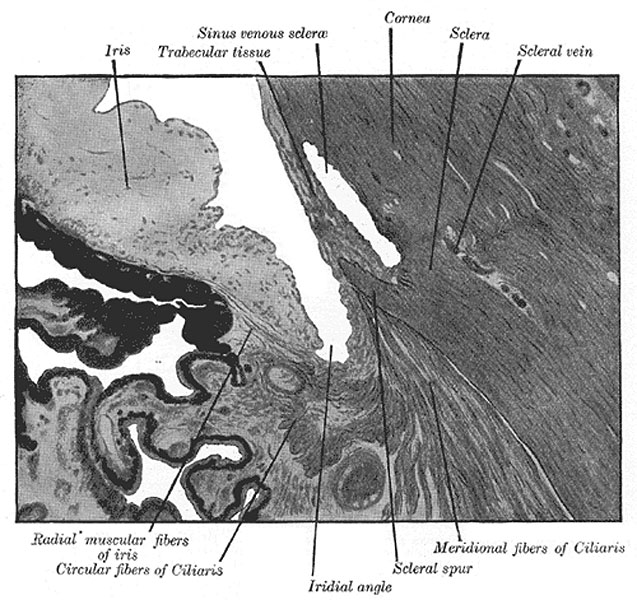
Cornea
Enlarged general view of the iridial angle. (Arthur Thomson.)
The cornea is the projecting transparent part of the external tunic, and forms the anterior sixth of the surface of the bulb. It is almost circular in outline, occasionally a little broader in the transverse than in the vertical direction. It is convex anteriorly and projects like a dome in front of the sclera. Its degree of curvature varies in different individuals, and in the same individual at different periods of life, being more pronounced in youth than in advanced life. The cornea is dense and of uniform thickness throughout; its posterior surface is perfectly circular in outline, and exceeds the anterior surface slightly in diameter. Immediately in front of the sclero-corneal junction the cornea bulges inward as a thickened rim, and behind this there is a distinct furrow between the attachment of the iris and the sclero-corneal junction. This furrow has been named by Arthur Thomson the sulcus circularis corneæ it is bounded externally by the trabecular tissue already described as forming the inner wall of the sinus venosus scleræ. Between this tissue and the anterior surface of the attached margin of the iris is an angular recess, named the iridial angle or filtration angle of the eye (Fig. 870).
(Text modified from Gray's 1918 Anatomy)
- Gray's Images: Development | Lymphatic | Neural | Vision | Hearing | Somatosensory | Integumentary | Respiratory | Gastrointestinal | Urogenital | Endocrine | Surface Anatomy | iBook | Historic Disclaimer
| Historic Disclaimer - information about historic embryology pages |
|---|
 |
| iBook - Gray's Embryology | |
|---|---|

|
|
Reference
Gray H. Anatomy of the human body. (1918) Philadelphia: Lea & Febiger.
Cite this page: Hill, M.A. (2024, April 27) Embryology Gray0870.jpg. Retrieved from https://embryology.med.unsw.edu.au/embryology/index.php/File:Gray0870.jpg
- © Dr Mark Hill 2024, UNSW Embryology ISBN: 978 0 7334 2609 4 - UNSW CRICOS Provider Code No. 00098G
File history
Click on a date/time to view the file as it appeared at that time.
| Date/Time | Thumbnail | Dimensions | User | Comment | |
|---|---|---|---|---|---|
| current | 09:11, 19 August 2012 | | 637 × 600 (109 KB) | Z8600021 (talk | contribs) | ==Cornea== Enlarged general view of the iridial angle. (Arthur Thomson.) The cornea is the projecting transparent part of the external tunic, and forms the anterior sixth of the surface of the bulb. It is almost circular in outline, occasionally a littl |
You cannot overwrite this file.
File usage
The following page uses this file: