File:Gray0869.jpg
Gray0869.jpg (748 × 600 pixels, file size: 126 KB, MIME type: image/jpeg)
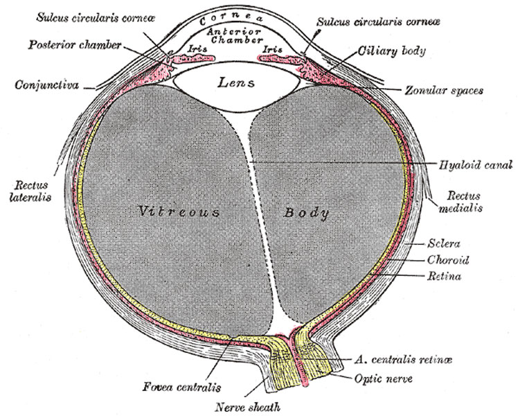
Horizontal section of the eyeball
The Sclera
The sclera has received its name from its extreme density and hardness; it is a firm, unyielding membrane, serving to maintain the form of the bulb. It is much thicker behind than in front; the thickness of its posterior part is 1 mm. Its external surface is of white color, and is in contact with the inner surface of the fascia of the bulb; it is quite smooth, except at the points where the Recti and Obliqui are inserted into it; its anterior part is covered by the conjunctival membrane. Its inner surface is brown in color and marked by grooves, in which the ciliary nerves and vessels are lodged; it is separated from the outer surface of the choroid by an extensive lymph space (spatium perichorioideale) which is traversed by an exceedingly fine cellular tissue, the lamina suprachorioidea. Behind it is pierced by the optic nerve, and is continuous through the fibrous sheath of this nerve with the dura mater. Where the optic nerve passes through the sclera, the latter forms a thin cribriform lamina, the lamina cribrosa scleræ; the minute orifices in this lamina serve for the transmission of the nervous filaments, and the fibrous septa dividing them from one another are continuous with the membranous processes which separate the bundles of nerve fibers. One of these openings, larger than the rest, occupies the center of the lamina; it transmits the central artery and vein of the retina. Around the entrance of the optic nerve are numerous small apertures for the transmission of the ciliary vessels and nerves, and about midway between this entrance and the sclerocorneal junction are four or five large apertures for the transmission of veins (venæ vorticosæ). In front, the sclera is directly continuous with the cornea, the line of union being termed the sclero-corneal junction. In the inner part of the sclera close to this junction is a circular canal, the sinus venosus scleræ (canal of Schlemm). In a meridional section of this region this sinus presents the appearance of a cleft, the outer wall of which consists of the firm tissue of the sclera, while its inner wall is formed by a triangular mass of trabecular tissue (Fig. 870); the apex of the mass is directed forward and is continuous with the posterior elastic lamina of the cornea. The sinus is lined by endothelium and communicates externally with the anterior ciliary veins.
Sclera Structure
The sclera is formed of white fibrous tissue intermixed with fine elastic fibers; flattened connective-tissue corpuscles, some of which are pigmented, are contained in cell spaces between the fibers. The fibers are aggregated into bundles, which are arranged chiefly in a longitudinal direction. Its vessels are not numerous, the capillaries being of small size, uniting at long and wide intervals. Its nerves are derived from the ciliary nerves, but their exact mode of ending is not known.
The Cornea
The cornea is the projecting transparent part of the external tunic, and forms the anterior sixth of the surface of the bulb. It is almost circular in outline, occasionally a little broader in the transverse than in the vertical direction. It is convex anteriorly and projects like a dome in front of the sclera. Its degree of curvature varies in different individuals, and in the same individual at different periods of life, being more pronounced in youth than in advanced life. The cornea is dense and of uniform thickness throughout; its posterior surface is perfectly circular in outline, and exceeds the anterior surface slightly in diameter. Immediately in front of the sclero-corneal junction the cornea bulges inward as a thickened rim, and behind this there is a distinct furrow between the attachment of the iris and the sclero-corneal junction. This furrow has been named by Arthur Thomson 145 the sulcus circularis corneæ it is bounded externally by the trabecular tissue already described as forming the inner wall of the sinus venosus scleræ. Between this tissue and the anterior surface of the attached margin of the iris is an angular recess, named the iridial angle or filtration angle of the eye (Fig. 870). Immediately outside the filtration angle is a projecting rim of scleral tissue which appears in a meridional section as a small triangular area, termed the scleral spur. Its base is continuous with the inner surface of the sclera immediately to the outer side of the filtration angle and its apex is directed forward and inward. To the anterior sloping margin of this spur are attached the bundles of trabecular tissue just referred to; from its posterior margin the meridional fibers of the Ciliaris muscle arise.
Cornea Structure
(Fig. 871) The cornea consists from before backward of four layers, viz.: (1) the corneal epithelium, continuous with that of the conjunctiva; (2) the substantia propria (3) the posterior elastic lamina; and (4) the endothelium of the anterior chamber. 7
 The corneal epithelium (epithelium corneæ anterior layer) covers the front of the cornea and consists of several layers of cells. The cells of the deepest layer are columnar; then follow two or three layers of polyhedral cells, the majority of which are prickle cells similar to those found in the stratum mucosum of the cuticle. Lastly, there are three or four layers of squamous cells, with flattened nuclei.	   8
 The substantia propria is fibrous, tough, unyielding, and perfectly transparent. It is composed of about sixty flattened lamellæ, superimposed one on another. These lamellæ are made up of bundles of modified connective tissue, the fibers of which are directly continuous with those of the sclera. The fibers of each lamella are for the most part parallel with one another, but at right angles to those of adjacent lamellæ. Fibers, however, frequently pass from one lamella to the next.
The lamellæ are connected with each other by an interstitial cement substance, in which are spaces, the corneal spaces. These are stellate in shape and communicate with one another by numerous offsets. Each contains a cell, the corneal corpuscle, resembling in form the space in which it is lodged, but not entirely filling it.
(Text modified from Gray's 1918 Anatomy)
- Gray's Images: Development | Lymphatic | Neural | Vision | Hearing | Somatosensory | Integumentary | Respiratory | Gastrointestinal | Urogenital | Endocrine | Surface Anatomy | iBook | Historic Disclaimer
| Historic Disclaimer - information about historic embryology pages |
|---|
| Pages where the terms "Historic" (textbooks, papers, people, recommendations) appear on this site, and sections within pages where this disclaimer appears, indicate that the content and scientific understanding are specific to the time of publication. This means that while some scientific descriptions are still accurate, the terminology and interpretation of the developmental mechanisms reflect the understanding at the time of original publication and those of the preceding periods, these terms, interpretations and recommendations may not reflect our current scientific understanding. (More? Embryology History | Historic Embryology Papers) |
| iBook - Gray's Embryology | |
|---|---|

|
|
Reference
Gray H. Anatomy of the human body. (1918) Philadelphia: Lea & Febiger.
Cite this page: Hill, M.A. (2024, April 27) Embryology Gray0869.jpg. Retrieved from https://embryology.med.unsw.edu.au/embryology/index.php/File:Gray0869.jpg
- © Dr Mark Hill 2024, UNSW Embryology ISBN: 978 0 7334 2609 4 - UNSW CRICOS Provider Code No. 00098G
File history
Click on a date/time to view the file as it appeared at that time.
| Date/Time | Thumbnail | Dimensions | User | Comment | |
|---|---|---|---|---|---|
| current | 09:08, 19 August 2012 |  | 748 × 600 (126 KB) | Z8600021 (talk | contribs) | ==Horizontal section of the eyeball== ===The Sclera=== The sclera has received its name from its extreme density and hardness; it is a firm, unyielding membrane, serving to maintain the form of the bulb. It is much thicker behind than in front; the thic |
You cannot overwrite this file.
File usage
The following page uses this file:

