File:Gray0616.jpg
Gray0616.jpg (626 × 600 pixels, file size: 111 KB, MIME type: image/jpeg)
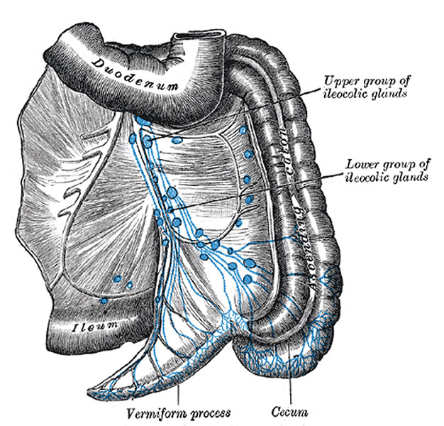
Lymphatics of Cecum and Vermiform Process
Viewed from behind. (Jamieson and Dobson.)
Mesenteric Glands (lymphoglandulæ mesentericæ) lie between the layers of the mesentery. They vary from one hundred to one hundred and fifty in number, and may be grouped into three sets, viz.: one lying close to the wall of the small intestine, among the terminal twigs of the superior mesenteric artery; a second, in relation to the loops and primary branches of the vessels; and a third along the trunk of the artery.
Ileocolic glands (Figs. 615, 616), from ten to twenty in number, form a chain around the ileocolic artery, but show a tendency to subdivision into two groups, one near the duodenum and another on the lower part of the trunk of the artery. Where the vessel divides into its terminal branches the chain is broken up into several groups:
- ileal, in relation to the ileal branch of the artery
- anterior ileocolic, usually of three glands, in the ileocolic fold, near the wall of the cecum
- posterior ileocolic, mostly placed in the angle between the ileum and the colon, but partly lying behind the cecum at its junction with the ascending colon
- a single gland, between the layers of the mesenteriole of the vermiform process
- right colic, along the medial side of the ascending colon.
(Text from Gray's Anatomy 1918)
- Gray's Images: Development | Lymphatic | Neural | Vision | Hearing | Somatosensory | Integumentary | Respiratory | Gastrointestinal | Urogenital | Endocrine | Surface Anatomy | iBook | Historic Disclaimer
| Historic Disclaimer - information about historic embryology pages |
|---|
 |
| iBook - Gray's Embryology | |
|---|---|

|
|
Reference
Gray H. Anatomy of the human body. (1918) Philadelphia: Lea & Febiger.
Cite this page: Hill, M.A. (2024, May 10) Embryology Gray0616.jpg. Retrieved from https://embryology.med.unsw.edu.au/embryology/index.php/File:Gray0616.jpg
- © Dr Mark Hill 2024, UNSW Embryology ISBN: 978 0 7334 2609 4 - UNSW CRICOS Provider Code No. 00098G
File history
Click on a date/time to view the file as it appeared at that time.
| Date/Time | Thumbnail | Dimensions | User | Comment | |
|---|---|---|---|---|---|
| current | 23:50, 14 February 2013 | | 626 × 600 (111 KB) | Z8600021 (talk | contribs) | ==Lymphatics of Cecum and Vermiform Process== Viewed from behind. (Jamieson and Dobson.) '''Mesenteric Glands''' (lymphoglandulæ mesentericæ) lie between the layers of the mesentery. They vary from one hundred to one hundred and fifty in number, and m |
You cannot overwrite this file.
File usage
The following 3 pages use this file: