File:Gray0602.jpg
Original file (700 × 708 pixels, file size: 132 KB, MIME type: image/jpeg)
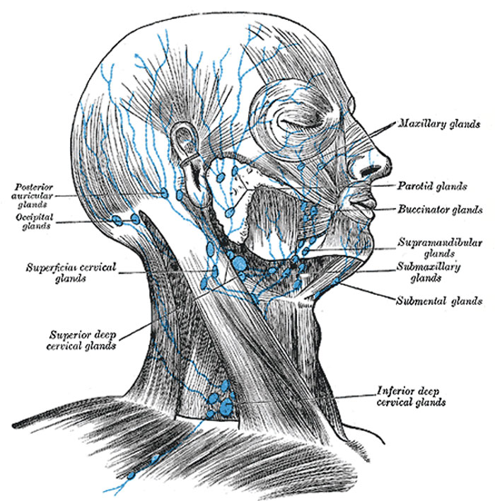
The Lymph Glands of the Head
Superficial lymph glands and lymphatic vessels of head and neck.
The lymph glands of the head are arranged in the following groups:
Occipital
|
Facial
|
Occipital glands
Occipital glands (lymphoglandulæ occipitales), one to three in number, are placed on the back of the head close to the margin of the Trapezius and resting on the insertion of the Semispinalis capitis. Their afferent vessels drain the occipital region of the scalp, while their efferents pass to the superior deep cervical glands.
Posterior auricular glands (lymphoglandulæ auriculares; mastoid glands), usually two in number, are situated on the mastoid insertion of the Sternocleidomastoideus, beneath the Auricularis posterior. Their afferent vessels drain the posterior part of the temporoparietal region, the upper part of the cranial surface of the auricula or pinna, and the back of the external acoustic meatus; their efferents pass to the superior deep cervical glands.
Anterior auricular glands (lymphoglandulæ auriculares anteriores; superficial parotid or preauricular glands), from one to three in number, lie immediately in front of the tragus. Their afferents drain the lateral surface of the auricula and the skin of the adjacent part of the temporal region; their efferents pass to the superior deep cervical glands.
Parotid glands (lymphoglandulæ parotideæ), form two groups in relation with the parotid salivary gland, viz., a group imbedded in the substance of the gland, and a group of subparotid glands lying on the lateral wall of the pharynx. Occasionally small glands are found in the subcutaneous tissue over the parotid gland. Their afferent vessels drain the root of the nose, the eyelids, the frontotemporal region, the external acoustic meatus and the tympanic cavity, possibly also the posterior parts of the palate and the floor of the nasal cavity. The efferents of these glands pass to the superior deep cervical glands. The afferents of the subparotid glands drain the nasal part of the pharynx and the posterior parts of the nasal cavities; their efferents pass to the superior deep cervical glands.
Facial glands
The facial glands comprise three groups: (a) infraorbital or maxillary, scattered over the infraorbital region from the groove between the nose and cheek to the zygomatic arch; (b) buccinator, one or more placed on the Buccinator opposite the angle of the mouth; (c) supramandibular, on the outer surface of the mandible, in front of the Masseter and in contact with the external maxillary artery and anterior facial vein. Their efferent vessels drain the eyelids, the conjunctiva, and the skin and mucous membrane of the nose and cheek; their efferents pass to the submaxillary glands.
Deep facial glands (lymphoglandulæ faciales profunda; internal maxillary glands) are placed beneath the ramus of the mandible, on the outer surface of the Pterygoideus externus, in relation to the internal maxillary artery. Their afferent vessels drain the temporal and infratemporal fossæ and the nasal part of the pharynx their efferents pass to the superior deep cervical glands.
Lingual glands (lymphoglandulæ linguales) are two or three small nodules lying on the Hyoglossus and under the Genioglossus. They form merely glandular substations in the course of the lymphatic vessels of the tongue.
Retropharyngeal glands (Fig. 603), from one to three in number, lie in the buccopharyngeal fascia, behind the upper part of the pharynx and in front of the arch of the atlas, being separated, however, from the latter by the Longus capitis. Their afferents drain the nasal cavities, the nasal part of the pharynx, and the auditory tubes; their efferents pass to the superior deep cervical glands.
(Text from Gray's Anatomy 1918)
Gray's Lymphatic Anatomy: 592 Primary lymph sacs | 593 Lymph capillaries of the human conjunctiva | 594 Lymph capillaries from the human scrotum | 595 Lymph capillaries of the sole of the human foot | 596 Section through human tongue | 597 Lymph gland (Node) | 598 Lymph gland tissue | 599 Thoracic and right lymphatic ducts | 600 Modes of origin of thoracic duct | 601 Terminal collecting trunks of right side | 602 Lymph glands of the head | 603 Lymphatics of pharynx | 604 Lymphatics of the face | 605 Lymphatics of the Tongue | 606 Lymph glands of the upper extremity | 607 Lymphatics of the mamma | 608 Lymphatic vessels of the dorsal hand surface | 609 Lymph glands of popliteal fossa | 610 Superficial lymph glands and vessels of the lower extremity | 611 Parietal lymph glands of the pelvis | 612 Iliopelvic glands | 613 Lymphatics of stomach | 614 Lymphatics of stomach | 615 Lymphatics of cecum and vermiform process | 616 Lymphatics of cecum and vermiform process | 617 Lymphatics of Colon | 618 Lymphatic of the Bladder | 619 Lymphatics of the Prostate | 620 Lymphatics of the Uterus | 621 Lymphatics of the thorax and abdomen | 622 Tracheobronchial Lymph Glands | Gray's Anatomy | Historic Disclaimer | Lymphatic Development
- Gray's Images: Development | Lymphatic | Neural | Vision | Hearing | Somatosensory | Integumentary | Respiratory | Gastrointestinal | Urogenital | Endocrine | Surface Anatomy | iBook | Historic Disclaimer
| Historic Disclaimer - information about historic embryology pages |
|---|
 |
| iBook - Gray's Embryology | |
|---|---|

|
|
Reference
Gray H. Anatomy of the human body. (1918) Philadelphia: Lea & Febiger.
Cite this page: Hill, M.A. (2024, April 27) Embryology Gray0602.jpg. Retrieved from https://embryology.med.unsw.edu.au/embryology/index.php/File:Gray0602.jpg
- © Dr Mark Hill 2024, UNSW Embryology ISBN: 978 0 7334 2609 4 - UNSW CRICOS Provider Code No. 00098G
File history
Click on a date/time to view the file as it appeared at that time.
| Date/Time | Thumbnail | Dimensions | User | Comment | |
|---|---|---|---|---|---|
| current | 15:34, 14 February 2013 | | 700 × 708 (132 KB) | Z8600021 (talk | contribs) | ==Head and Neck Lymph Glands and Vessels== Superficial lymph glands and lymphatic vessels of head and neck. (Text from Gray's Anatomy 1918) {{Gray Anatomy}} Category:Immune |
You cannot overwrite this file.
File usage
The following 3 pages use this file: