File:Gray0601.jpg
Original file (1,000 × 357 pixels, file size: 62 KB, MIME type: image/jpeg)
The Thoracic and Right Lymphatic Ducts
Thoracic Duct
The thoracic duct (ductus thoracicus) (Fig. 599) conveys the greater part of the lymph and chyle into the blood. It is the common trunk of all the lymphatic vessels of the body, excepting those on the right side of the head, neck, and thorax, and right upper extremity, the right lung, right side of the heart, and the convex surface of the liver. In the adult it varies in length from 38 to 45 cm. and extends from the second lumbar vertebra to the root of the neck.
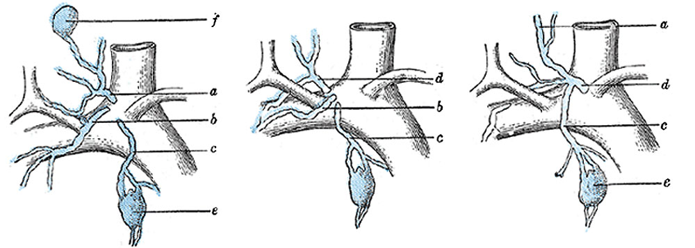
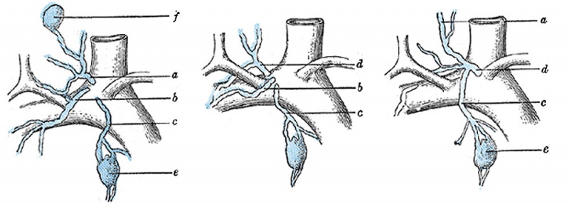
Right Lymphatic Duct
The right lymphatic duct (ductus lymphaticus dexter) (Fig. 601), about 1.25 cm. in length, courses along the medial border of the Scalenus anterior at the root of the neck and ends in the right subclavian vein, at its angle of junction with the right internal jugular vein. Its orifice is guarded by two semilunar valves, which prevent the passage of venous blood into the duct.
- Gray's Images: Development | Lymphatic | Neural | Vision | Hearing | Somatosensory | Integumentary | Respiratory | Gastrointestinal | Urogenital | Endocrine | Surface Anatomy | iBook | Historic Disclaimer
| Historic Disclaimer - information about historic embryology pages |
|---|
 |
| iBook - Gray's Embryology | |
|---|---|

|
|
Reference
Gray H. Anatomy of the human body. (1918) Philadelphia: Lea & Febiger.
Cite this page: Hill, M.A. (2024, May 21) Embryology Gray0601.jpg. Retrieved from https://embryology.med.unsw.edu.au/embryology/index.php/File:Gray0601.jpg
- © Dr Mark Hill 2024, UNSW Embryology ISBN: 978 0 7334 2609 4 - UNSW CRICOS Provider Code No. 00098G
File history
Click on a date/time to view the file as it appeared at that time.
| Date/Time | Thumbnail | Dimensions | User | Comment | |
|---|---|---|---|---|---|
| current | 14:54, 14 February 2013 | 1,000 × 357 (62 KB) | Z8600021 (talk | contribs) |
You cannot overwrite this file.
File usage
The following 3 pages use this file: