File:Gray0601.jpg: Difference between revisions
(uploaded a new version of "File:Gray0601.jpg") |
No edit summary |
||
| Line 1: | Line 1: | ||
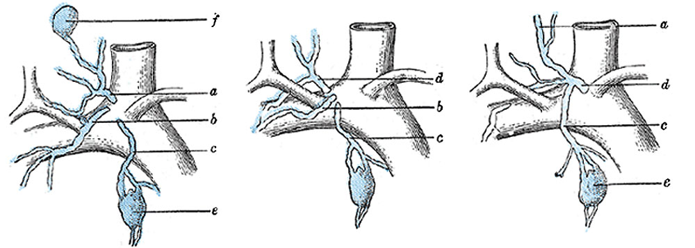
== | ==Terminal Collecting Trunks of Right Side== | ||
(Poirier and Charpy.) | (Poirier and Charpy.) | ||
{| | {| | ||
| '''Legend''' | | width=250px|'''Legend''' | ||
* a. | * a. Jugular trunk. | ||
* | * b. Subclavian trunk. | ||
* | * c. Bronchomediastinal trunk. | ||
* d. | * d. Right lymphatic trunk. | ||
* e. | * e. Gland of internal mammary chain. | ||
* | * f. Gland of deep cervical chain. (Poirier and Charpy.) | ||
| | | | ||
'''Right lymphatic duct''' - (ductus lymphaticus dexter) ([[:File:Gray0601.jpg|Fig. 601]]), about 1.25 cm. in length, courses along the medial border of the Scalenus anterior at the root of the neck and ends in the right subclavian vein, at its angle of junction with the right internal jugular vein. Its orifice is guarded by two semilunar valves, which prevent the passage of venous blood into the duct. | '''Right lymphatic duct''' - (ductus lymphaticus dexter) ([[:File:Gray0601.jpg|Fig. 601]]), about 1.25 cm. in length, courses along the medial border of the Scalenus anterior at the root of the neck and ends in the right subclavian vein, at its angle of junction with the right internal jugular vein. Its orifice is guarded by two semilunar valves, which prevent the passage of venous blood into the duct. | ||
|} | |} | ||
Revision as of 14:56, 14 February 2013
Terminal Collecting Trunks of Right Side
(Poirier and Charpy.)
Legend
|
Right lymphatic duct - (ductus lymphaticus dexter) (Fig. 601), about 1.25 cm. in length, courses along the medial border of the Scalenus anterior at the root of the neck and ends in the right subclavian vein, at its angle of junction with the right internal jugular vein. Its orifice is guarded by two semilunar valves, which prevent the passage of venous blood into the duct. |
- Gray's Images: Development | Lymphatic | Neural | Vision | Hearing | Somatosensory | Integumentary | Respiratory | Gastrointestinal | Urogenital | Endocrine | Surface Anatomy | iBook | Historic Disclaimer
| Historic Disclaimer - information about historic embryology pages |
|---|
 |
| iBook - Gray's Embryology | |
|---|---|

|
|
Reference
Gray H. Anatomy of the human body. (1918) Philadelphia: Lea & Febiger.
Cite this page: Hill, M.A. (2024, May 22) Embryology Gray0601.jpg. Retrieved from https://embryology.med.unsw.edu.au/embryology/index.php/File:Gray0601.jpg
- © Dr Mark Hill 2024, UNSW Embryology ISBN: 978 0 7334 2609 4 - UNSW CRICOS Provider Code No. 00098G
File history
Click on a date/time to view the file as it appeared at that time.
| Date/Time | Thumbnail | Dimensions | User | Comment | |
|---|---|---|---|---|---|
| current | 14:54, 14 February 2013 | 1,000 × 357 (62 KB) | Z8600021 (talk | contribs) |
You cannot overwrite this file.
File usage
The following 3 pages use this file: