File:Gray0476.jpg
From Embryology

Size of this preview: 591 × 600 pixels. Other resolution: 600 × 609 pixels.
Original file (600 × 609 pixels, file size: 99 KB, MIME type: image/jpeg)
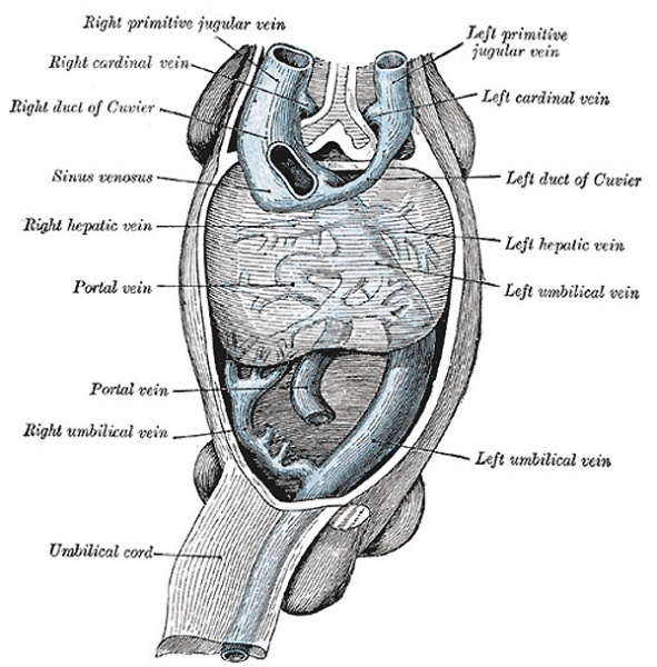
Human Sinus Venosus and its Tributaries
Human embryo with heart and anterior body-wall removed to show the sinus venosus and its tributaries.
(After His.)
- The right umbilical and right vitelline veins shrivel and disappear
- the left umbilical becomes enlarged and opens into the upper venous ring of the vitelline veins
- with the atrophy of the yolk-sac the left vitelline vein also undergoes atrophy and disappears
- a direct branch is established between this ring and the right hepatic vein
- this branch is named the ductus venosus
ductus venosus
- at the porta hepatis (transverse fissure of the liver) the umbilical vein divides into two branches
- the larger is joined by the portal vein, and enters the right lobe
- the smaller is continued upward (ductus venosus) and joins the inferior vena cava
- forms a wide channel
- blood returned from the placenta is carried directly to the heart without passing through the liver
- Gray's Images: Development | Lymphatic | Neural | Vision | Hearing | Somatosensory | Integumentary | Respiratory | Gastrointestinal | Urogenital | Endocrine | Surface Anatomy | iBook | Historic Disclaimer
| Historic Disclaimer - information about historic embryology pages |
|---|
| Pages where the terms "Historic" (textbooks, papers, people, recommendations) appear on this site, and sections within pages where this disclaimer appears, indicate that the content and scientific understanding are specific to the time of publication. This means that while some scientific descriptions are still accurate, the terminology and interpretation of the developmental mechanisms reflect the understanding at the time of original publication and those of the preceding periods, these terms, interpretations and recommendations may not reflect our current scientific understanding. (More? Embryology History | Historic Embryology Papers) |
| iBook - Gray's Embryology | |
|---|---|

|
|
Reference
Gray H. Anatomy of the human body. (1918) Philadelphia: Lea & Febiger.
Cite this page: Hill, M.A. (2024, April 27) Embryology Gray0476.jpg. Retrieved from https://embryology.med.unsw.edu.au/embryology/index.php/File:Gray0476.jpg
- © Dr Mark Hill 2024, UNSW Embryology ISBN: 978 0 7334 2609 4 - UNSW CRICOS Provider Code No. 00098G
File history
Click on a date/time to view the file as it appeared at that time.
| Date/Time | Thumbnail | Dimensions | User | Comment | |
|---|---|---|---|---|---|
| current | 11:19, 6 May 2011 |  | 600 × 609 (99 KB) | S8600021 (talk | contribs) | |
| 11:18, 6 May 2011 |  | 600 × 609 (63 KB) | S8600021 (talk | contribs) | ||
| 11:17, 6 May 2011 |  | 600 × 609 (53 KB) | S8600021 (talk | contribs) | ||
| 22:25, 11 October 2009 |  | 493 × 500 (44 KB) | S8600021 (talk | contribs) | Human embryo with heart and anterior body-wall removed to show the sinus venosus and its tributaries. (After His.) |
You cannot overwrite this file.
File usage
The following 3 pages use this file:
